Copyright
©2007 Baishideng Publishing Group Co.
World J Gastroenterol. Jan 21, 2007; 13(3): 480-482
Published online Jan 21, 2007. doi: 10.3748/wjg.v13.i3.480
Published online Jan 21, 2007. doi: 10.3748/wjg.v13.i3.480
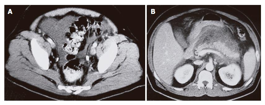
Figure 1 CT of the abdomen with contrast showing pelvic ascites (A) and inflammatory changes surrounding the pancreas (B).
The pancreas is well demarcated and homogenous with no focal lesions.
- Citation: Khan FY, Matar I. Chylous ascites secondary to hyperlipidemic pancreatitis with normal serum amylase and lipase. World J Gastroenterol 2007; 13(3): 480-482
- URL: https://www.wjgnet.com/1007-9327/full/v13/i3/480.htm
- DOI: https://dx.doi.org/10.3748/wjg.v13.i3.480


