Copyright
©2007 Baishideng Publishing Group Co.
World J Gastroenterol. Jan 21, 2007; 13(3): 426-431
Published online Jan 21, 2007. doi: 10.3748/wjg.v13.i3.426
Published online Jan 21, 2007. doi: 10.3748/wjg.v13.i3.426
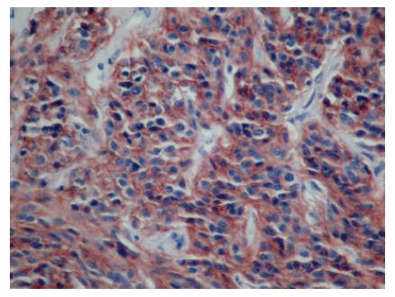
Figure 1 Diffuse cytoplasmic staining of COX-2 in the tumor cells of GIST.
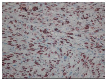
Figure 2 High PCNA immunoreactivity in GIST with more than 50% of nuclear positivity.
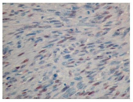
Figure 3 Low Ki-67 immunoreactivity in GIST with less than 50% of nuclear positivity.
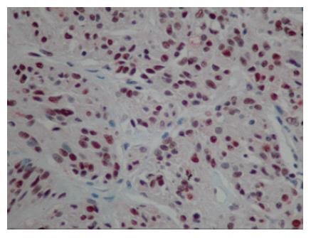
Figure 4 High p53 immunoreactivity in GIST with more than 75% of nuclear positivity.
- Citation: Gumurdulu D, Erdogan S, Kayaselcuk F, Seydaoglu G, Parsak CK, Demircan O, Tuncer I. Expression of COX-2, PCNA, Ki-67 and p53 in gastrointestinal stromal tumors and its relationship with histopathological parameters. World J Gastroenterol 2007; 13(3): 426-431
- URL: https://www.wjgnet.com/1007-9327/full/v13/i3/426.htm
- DOI: https://dx.doi.org/10.3748/wjg.v13.i3.426


