Copyright
©2007 Baishideng Publishing Group Co.
World J Gastroenterol. Aug 7, 2007; 13(29): 4027-4029
Published online Aug 7, 2007. doi: 10.3748/wjg.v13.i29.4027
Published online Aug 7, 2007. doi: 10.3748/wjg.v13.i29.4027
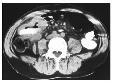
Figure 1 An abdominal CT scan section showing an obstructive luminal mass at the jejunum proved to be of melanotic nature.
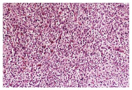
Figure 2 Melanoma of the small intestine with submucosal infiltration.
Numerous pleomorphic tumor cells with melanin deposits are viewed (HE, x 250).
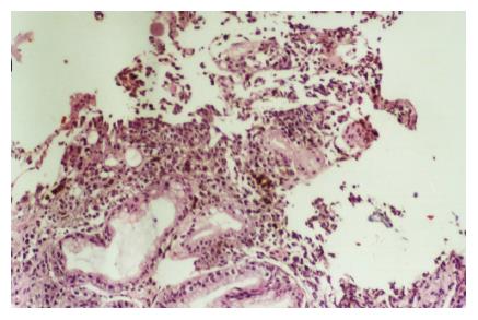
Figure 3 Colonic malignant melanoma originated at the anorectum (HE, x 100).
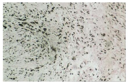
Figure 4 Melanoma of the small intestine stained with Masson-Fontana.
Plenty of granules of melanin are viewed (x 100).
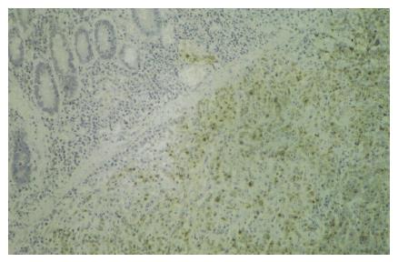
Figure 5 Melanoma of the small intestine stained with HMB-45 (x 100).
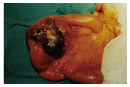
Figure 6 Surgical specimen of a segmental enterectomy for a primary melanoma located at the jejunum.
A few lymph nodes of the mesentery proved to be infiltrated.
- Citation: Manouras A, Genetzakis M, Lagoudianakis E, Markogiannakis H, Papadima A, Kafiri G, Filis K, Kekis P, Katergiannakis V. Malignant gastrointestinal melanomas of unknown origin: Should it be considered primary? World J Gastroenterol 2007; 13(29): 4027-4029
- URL: https://www.wjgnet.com/1007-9327/full/v13/i29/4027.htm
- DOI: https://dx.doi.org/10.3748/wjg.v13.i29.4027


