Copyright
©2007 Baishideng Publishing Group Co.
World J Gastroenterol. Aug 7, 2007; 13(29): 3973-3976
Published online Aug 7, 2007. doi: 10.3748/wjg.v13.i29.3973
Published online Aug 7, 2007. doi: 10.3748/wjg.v13.i29.3973
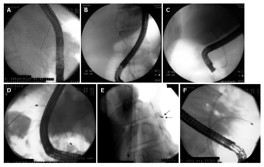
Figure 1 A: The papilla identified and cannulated by a papillotome and a guide wire is inserted deeply into the biliary tree by fluoroscopy; B: Contrast-free guidewire cannulation of main biliary duct (case of pancreatic cancer); C: Contrast-free deployment of plastic stent (same case of A); D: Contrast-free guidewire cannulation of left hepatic duct (case of Klatskin tumor); E: Contrast-free unilateral deployment of plastic stent (same case of C).
The arrows show the markers of the stent; F: Contrast-free bilateral deployment of plastic stent (case of Klatskin tumor).
- Citation: De Palma GD, Lombardi G, Rega M, Simeoli I, Masone S, Siciliano S, Maione F, Salvatori F, Balzano A, Persico G. Contrast-free endoscopic stent insertion in malignant biliary obstruction. World J Gastroenterol 2007; 13(29): 3973-3976
- URL: https://www.wjgnet.com/1007-9327/full/v13/i29/3973.htm
- DOI: https://dx.doi.org/10.3748/wjg.v13.i29.3973


