Copyright
©2007 Baishideng Publishing Group Co.
World J Gastroenterol. Jul 28, 2007; 13(28): 3897-3899
Published online Jul 28, 2007. doi: 10.3748/wjg.v13.i28.3897
Published online Jul 28, 2007. doi: 10.3748/wjg.v13.i28.3897
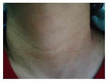
Figure 1 View of the patient’s skin from neck region.
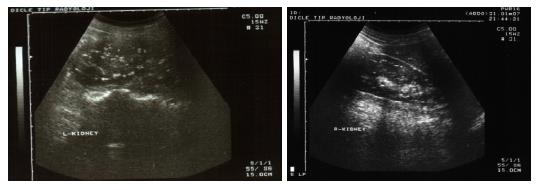
Figure 2 Nephrocalcinosis findings in both of the kidneys.
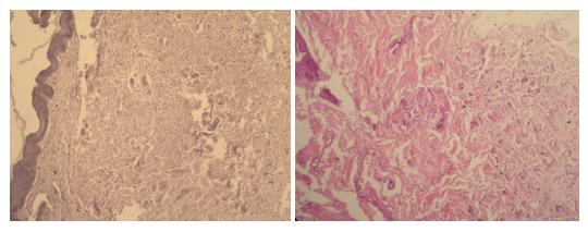
Figure 3 Degeneration in reticular fibers of dermis (HE).
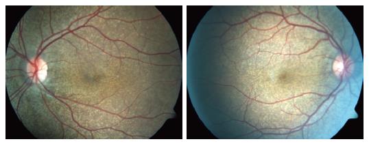
Figure 4 An appearance consistent of peau d'orange sign, peripapillary atrophy, and angioid streak in both of the eyes.
- Citation: Goral V, Demir D, Tuzun Y, Keklikci U, Buyukbayram H, Bayan K, Uyar A. Pseudoxantoma elasticum, as a repetitive upper gastrointestinal hemorrhage cause in a pregnant woman. World J Gastroenterol 2007; 13(28): 3897-3899
- URL: https://www.wjgnet.com/1007-9327/full/v13/i28/3897.htm
- DOI: https://dx.doi.org/10.3748/wjg.v13.i28.3897


