Copyright
©2007 Baishideng Publishing Group Inc.
World J Gastroenterol. Jul 14, 2007; 13(26): 3610-3613
Published online Jul 14, 2007. doi: 10.3748/wjg.v13.i26.3610
Published online Jul 14, 2007. doi: 10.3748/wjg.v13.i26.3610
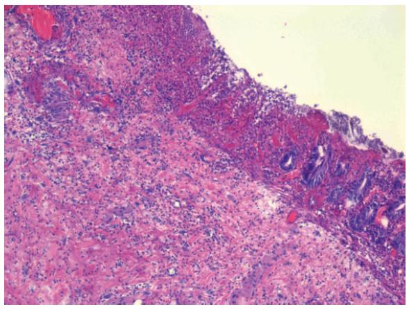
Figure 1 Photomicrograph of colonic resection specimen, taken with a light microscope (HE, x 10).
The image shows mucosa and submucosa. There is partial mucosal necrosis with significant loss of tissue architecture in the mucosa. In the lower right part of the picture, the mucosa is more intact. The submucosa shows fibrosis and a patchy chronic inflammation in the form of aggregates of lymphocytes.
- Citation: Larsen A, Reitan JB, Aase ST, Hauer-Jensen M. Long-term prognosis in patients with severe late radiation enteropathy: A prospective cohort study. World J Gastroenterol 2007; 13(26): 3610-3613
- URL: https://www.wjgnet.com/1007-9327/full/v13/i26/3610.htm
- DOI: https://dx.doi.org/10.3748/wjg.v13.i26.3610


