Copyright
©2007 Baishideng Publishing Group Inc.
World J Gastroenterol. Jul 14, 2007; 13(26): 3575-3580
Published online Jul 14, 2007. doi: 10.3748/wjg.v13.i26.3575
Published online Jul 14, 2007. doi: 10.3748/wjg.v13.i26.3575
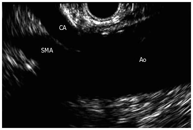
Figure 1 Linear EUS images of the aorta (Ao), celiac axis (CA), and superior mesenteric artery (SMA).
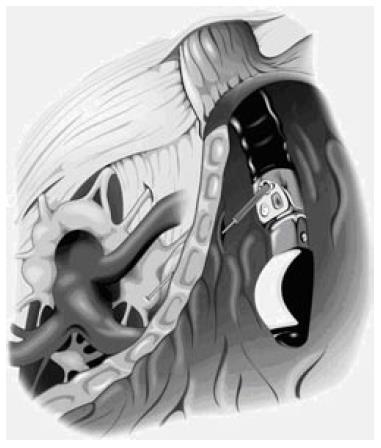
Figure 2 Illustration of the position of the celiac plexus, celiac trunk, and stomach when performing EUS-guided celiac injection.
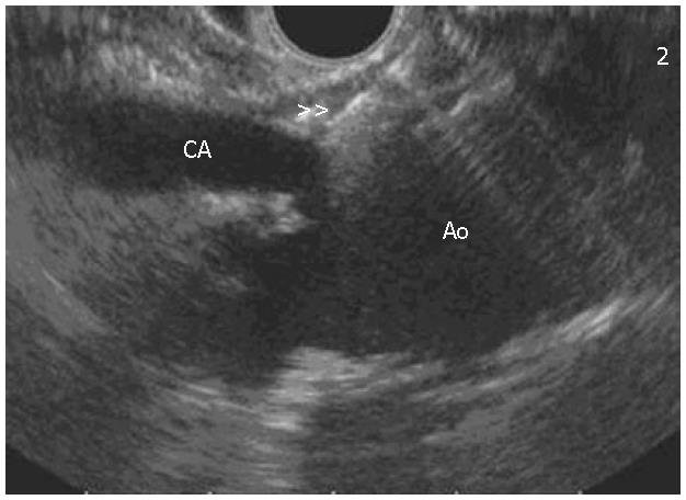
Figure 3 EUS image of the needle (double arrows) located at the celiac plexus in relation to the celiac axis (CA) and aorta (Ao).
- Citation: Michaels AJ, Draganov PV. Endoscopic ultrasonography guided celiac plexus neurolysis and celiac plexus block in the management of pain due to pancreatic cancer and chronic pancreatitis. World J Gastroenterol 2007; 13(26): 3575-3580
- URL: https://www.wjgnet.com/1007-9327/full/v13/i26/3575.htm
- DOI: https://dx.doi.org/10.3748/wjg.v13.i26.3575


