Copyright
©2007 Baishideng Publishing Group Co.
World J Gastroenterol. Jun 28, 2007; 13(24): 3301-3310
Published online Jun 28, 2007. doi: 10.3748/wjg.v13.i24.3301
Published online Jun 28, 2007. doi: 10.3748/wjg.v13.i24.3301
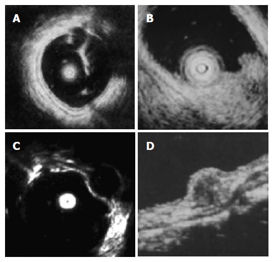
Figure 1 Endoscopic ultrasonography imagines of normal wall and submucosal tumors of the large intestine are presented.
A: The normal wall displayed in 5 layers; B: Lipoma imagine showing a hyperechoic homogeneous mass located in the third layer; C: Leiomyoma imagine showing a hypoechoic homogeneous mass originated from the 4th layer; D: Rectal carcinoid imagine showing a submucosal hypoechoic mass with a homogenous echo. Courtesy by PH Zhou (Zhou, 2004 128 /id).
- Citation: Ponsaing LG, Kiss K, Loft A, Jensen LI, Hansen MB. Diagnostic procedures for submucosal tumors in the gastrointestinal tract. World J Gastroenterol 2007; 13(24): 3301-3310
- URL: https://www.wjgnet.com/1007-9327/full/v13/i24/3301.htm
- DOI: https://dx.doi.org/10.3748/wjg.v13.i24.3301


