Copyright
©2007 Baishideng Publishing Group Co.
World J Gastroenterol. Jun 21, 2007; 13(23): 3189-3198
Published online Jun 21, 2007. doi: 10.3748/wjg.v13.i23.3189
Published online Jun 21, 2007. doi: 10.3748/wjg.v13.i23.3189
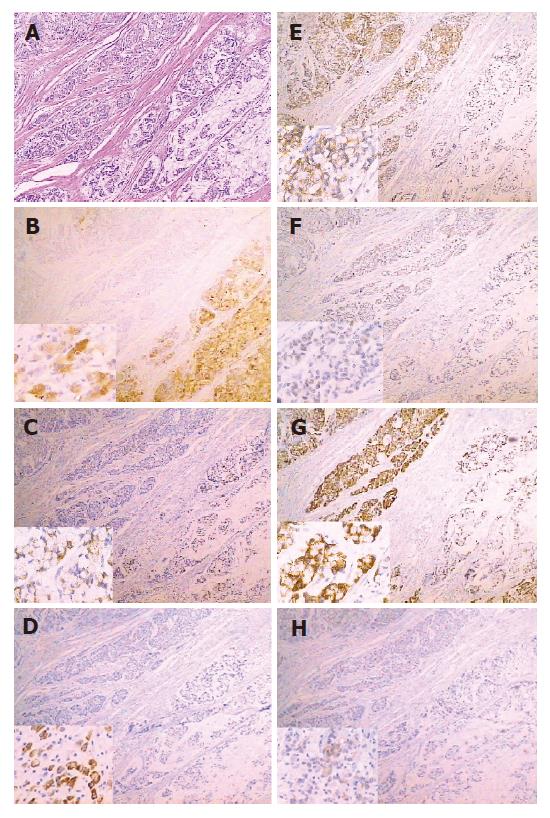
Figure 1 A case of signet ring gastric cancer of GI type showed MUC5AC (+), HGM (+), MUC6 (-), Li-cadherin (+), CDX2 (+), MUC2 (+) and VILLIN (-).
A: HE × 40; B: MUC5AC; C: HGM; D: MUC6; E: Li-cadherin; F: CDX2; G: MUC2; H: VILLIN, (B-H: Background picture is immunohistochemical staining of this case by each marker, × 40; left and lower pictures show positive staining of each marker, × 400).
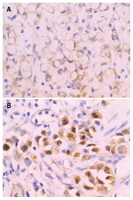
Figure 2 A: β-catenin membrane expression in a G type case (β-catenin staining, × 400); B: β-catenin plasma/nuclues expression in a GI type case (β-catenin staining, × 400).
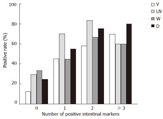
Figure 3 Clinicopathological features of different intestinal marker expression patterns.
V: vascular invasion; LN: lymph node metastasis; W: wall invasion deeper than submucosa layer; D: tumor diameter larger than 5 cm. The number of positive intestinal phenotype markers in one case was positively correlated with higher rates of lymph node metastasis (P < 0.01), vascular invasion (P < 0.01), larger tumor diameter (P < 0.01) and deeper wall invasion (P < 0.05). (Spearman's rank correlation analysis).
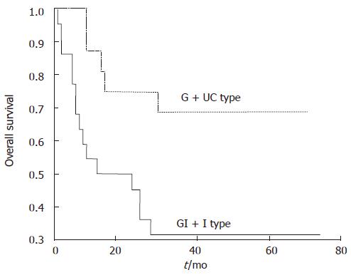
Figure 4 Gastric SRC carcinomas expressing intestinal phenotype markers (GI+I type cases) had significantly lower survival rates than those with no expression of intestinal phenotype markers (G + UC type cases), P = 0.
0146 by Kaplan-Meier method.
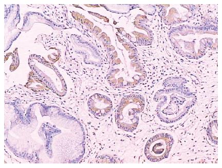
Figure 5 Li-cadherin expression in intestinal metapalsia of gastric mucosa (Li-cadherin staining, × 100).
- Citation: Tian MM, Zhao AL, Li ZW, Li JY. Phenotypic classification of gastric signet ring cell carcinoma and its relationship with clinicopathologic parameters and prognosis. World J Gastroenterol 2007; 13(23): 3189-3198
- URL: https://www.wjgnet.com/1007-9327/full/v13/i23/3189.htm
- DOI: https://dx.doi.org/10.3748/wjg.v13.i23.3189


