Copyright
©2007 Baishideng Publishing Group Co.
World J Gastroenterol. Jun 21, 2007; 13(23): 3153-3158
Published online Jun 21, 2007. doi: 10.3748/wjg.v13.i23.3153
Published online Jun 21, 2007. doi: 10.3748/wjg.v13.i23.3153
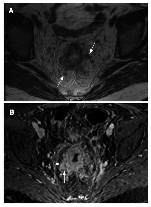
Figure 1 A 52 years old woman with rectal cancer.
Axial T2 (A) and axial fat suppressed gadolinium-enhanced T1-weighted (B) MR images demonstrate circumferential soft tissue thickening and abnormal enhancement of the rectum consistent with a neoplasm. Direct mesorectal invasion is present (arrows in B) as well as perirectal adenopathy (arrows in A).
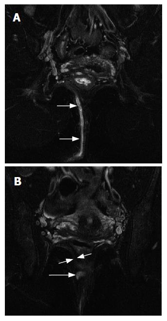
Figure 2 A 34 year old woman with a transphincteric peri-anal fistula.
Coronal fat suppressed T2-weighted MR images demonstrate a long right sided peri-anal fistula (arrows in A) which drains to the right gluteal cleft. In B, notice the fistulous tract (long arrow) extending through the levator muscle (short arrows). There is no evidence of abscess along the tract.
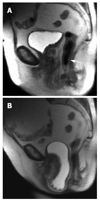
Figure 3 A 55 years old woman with a pelvic floor relaxation.
A: Sagittal T2-weighted MR image obtained at rest demonstrates a rectocele (arrow); B: Sagittal T2-weighted MR image obtained during a Valsalva maneuver demonstrates a severe cystocele (arrow) which impinges on the anterior wall of the rectum.
- Citation: Berman L, Israel GM, McCarthy SM, Weinreb JC, Longo WE. Utility of magnetic resonance imaging in anorectal disease. World J Gastroenterol 2007; 13(23): 3153-3158
- URL: https://www.wjgnet.com/1007-9327/full/v13/i23/3153.htm
- DOI: https://dx.doi.org/10.3748/wjg.v13.i23.3153


