Copyright
©2007 Baishideng Publishing Group Co.
World J Gastroenterol. Jun 14, 2007; 13(22): 3128-3132
Published online Jun 14, 2007. doi: 10.3748/wjg.v13.i22.3128
Published online Jun 14, 2007. doi: 10.3748/wjg.v13.i22.3128
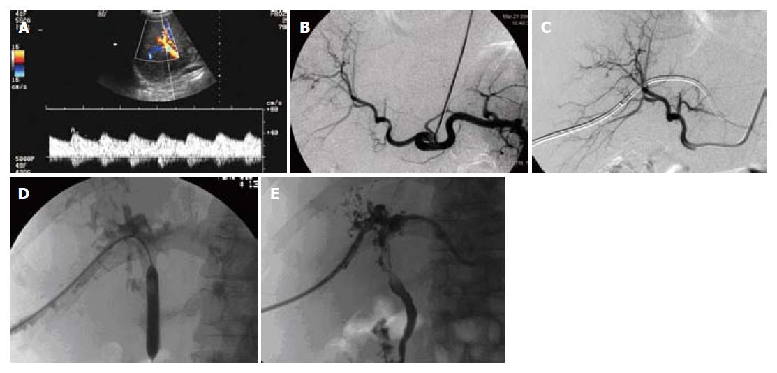
Figure 1 Images pertaining to case 2.
HA stoma stricture was diagnosed 3 mo after OLT, no treatment was performed; coronary artery stent placement was performed after 5 mo. Diffuse intra- and extra- hepatic bile duct stricture was apparent 8 mo after OLT. PTBD and balloon dilation therapy were performed. A: Tardus parvus frequency spectrum shown by color Doppler ultrasonography indicating HA stoma stricture; B: HA stoma stricture as indicated by celiac axis angiography; C: Celiac axis angiography revealing HAS disappearance following HA stent placement; D: Treatment of bile duct stricture by percutaneous transhepatic balloon dilation; E: Cholangiography revealed bile duct stiffness, rare branching, and filling defect in the bile duct lumina.
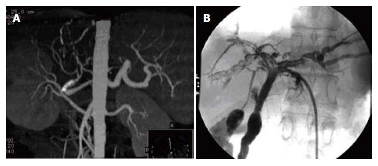
Figure 2 Images pertaining to case 3.
HAS was diagnosed 4 d after OLT, and coronary artery stent placement was performed. Diffuse intra- and extra-hepatic bile duct stricture was apparent 10 mo after OLT. PTBD, balloon dilation and bile duct stent placement therapy were performed. A: Computed tomography angiography revealing HAS disappearance following HA stent placement; B: Cholangiography showing bile duct stiffness, rare branching, and filling defect in the bile duct lumina.
- Citation: Zhao DB, Shan H, Jiang ZB, Huang MS, Zhu KS, Chen GH, Meng XC, Guan SH, Li ZR, Qian JS. Role of interventional therapy in hepatic artery stenosis and non-anastomosis bile duct stricture after orthotopic liver transplantation. World J Gastroenterol 2007; 13(22): 3128-3132
- URL: https://www.wjgnet.com/1007-9327/full/v13/i22/3128.htm
- DOI: https://dx.doi.org/10.3748/wjg.v13.i22.3128


