Copyright
©2007 Baishideng Publishing Group Co.
World J Gastroenterol. May 28, 2007; 13(20): 2803-2810
Published online May 28, 2007. doi: 10.3748/wjg.v13.i20.2803
Published online May 28, 2007. doi: 10.3748/wjg.v13.i20.2803
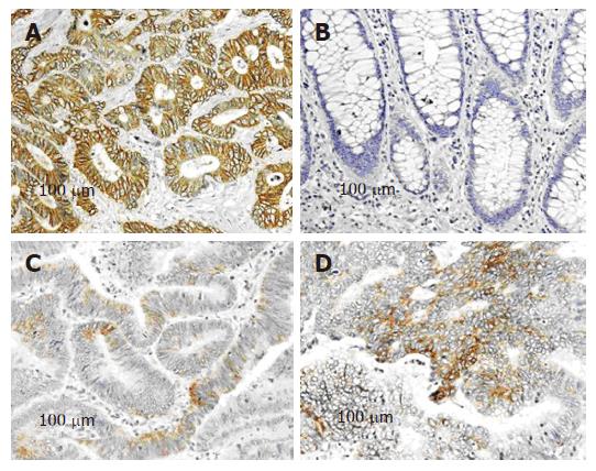
Figure 1 Expression of NDRG1 in colorectal cancer as analyzed by immunohistochemistry.
A: Moderately differentiated colorectal adenocarcinoma from a Japanese patient demonstrating membranous and cytoplasmic staining with NDRG1 antibody; B: No NDRG1 expression was detected in normal adjacent colon mucosa; C: Well differentiated colorectal adenocarcinoma from US patient; D: Moderately differentiated colorectal adenocarcinoma form US patient.
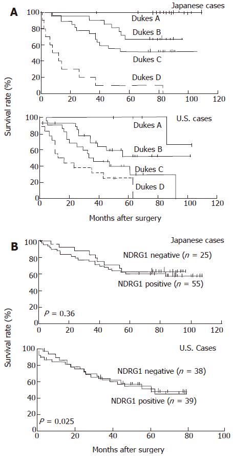
Figure 2 Kaplan-Meier survival analysis of colorectal cancer patients.
A: Survival (months after surgery) of Japanese and US patients stratified according to Dukes’ staging; B: Correlation of NDRG1 expression (negative, 0; positive, +1 to +3) with survival in Japanese patients and US patients (including Caucasians and African Americans).
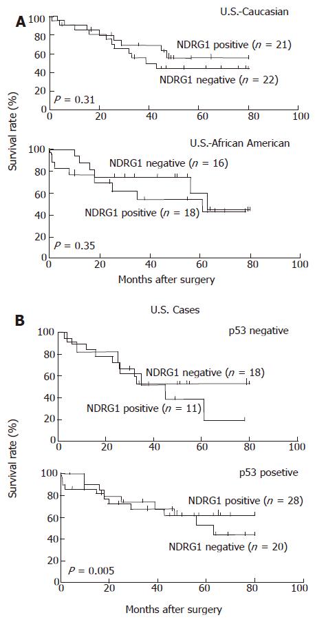
Figure 3 Kaplan-Meier survival analysis of US colorectal cancer patients according to NDRG1 expression.
A: Comparison of survival according to race/ethnicity, i.e., Caucasian and African American; B: Correlation of survival in p53-negative and p53-positive staining tumors.
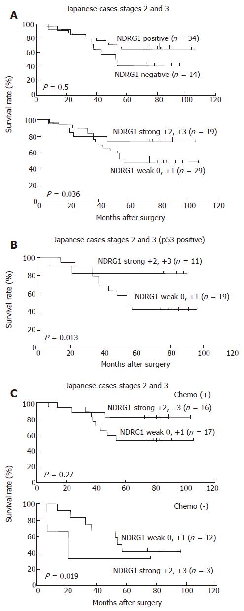
Figure 4 Kaplan-Meier survival analysis of NDRG1 expression in Japanese patients with stages II and III colorectal cancers.
A: Correlation of survival with NDRG1 expression stratified as negative (0) or positive (+1 to +3) and weak (0, +1) or strong (+2, +3); B: Correlation of survival with weak or strong NDRG1 staining in p53-positive tumors; C: Correlation of survival with weak or strong NDRG1 staining in patients with or without chemotherapeutic treatment, chemo (+) or chemo (-), respectively.
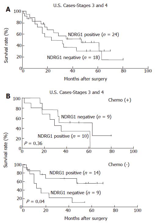
Figure 5 Kaplan-Meier survival analysis of NDRG1 expression in US with stages III and IV colorectal cancers.
A: Correlation of survival with NDRG1 expression stratified as negative (0) or positive (+1 to +3); B: Correlation of survival with weak or strong NDRG1 staining in patients with or without chemotherapeutic treatment, chemo (+) or chemo (-), respectively.
- Citation: Koshiji M, Kumamoto K, Morimura K, Utsumi Y, Aizawa M, Hoshino M, Ohki S, Takenoshita S, Costa M, Commes T, Piquemal D, Harris CC, Tchou-Wong KM. Correlation of N-myc downstream-regulated gene 1 expression with clinical outcomes of colorectal cancer patients of different race/ethnicity. World J Gastroenterol 2007; 13(20): 2803-2810
- URL: https://www.wjgnet.com/1007-9327/full/v13/i20/2803.htm
- DOI: https://dx.doi.org/10.3748/wjg.v13.i20.2803


