Copyright
©2007 Baishideng Publishing Group Co.
World J Gastroenterol. May 21, 2007; 13(19): 2758-2760
Published online May 21, 2007. doi: 10.3748/wjg.v13.i19.2758
Published online May 21, 2007. doi: 10.3748/wjg.v13.i19.2758
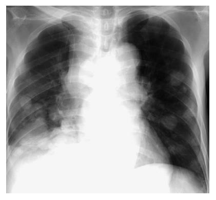
Figure 1 Chest X-ray.
Many nodules are present in the bilateral lung fields, and the mediastinal lymph node is enlarged.
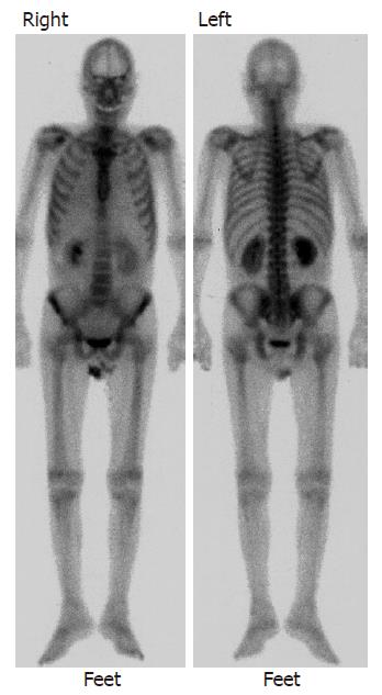
Figure 2 Bone scintigraphy.
No abnormal accumulation suggestive of metastasis is noted in the whole body bone.
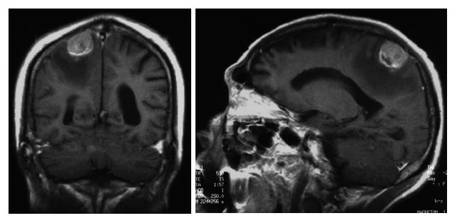
Figure 3 Head MRI.
A 2.5-cm tumor heterogeneously enhanced on contrast imaging in the parietal region. A low-intensity area, is an edematous change, extended from the right parietal region to the frontal lobe white matter.
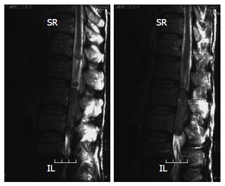
Figure 4 Spinal cord MRI.
Tumorous lesions are diffusely present from medullary cone to cauda equina, and located mostly in the spinal canal at the middle to lower lumbar level. The cauda equina is severely compressed.
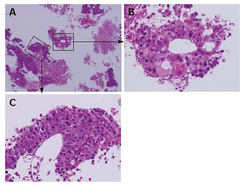
Figure 5 A: Pathology.
Atypical cells with granular eosinophilic cytoplasm, forming pseudoglandular and trabecular structures, suggested metastatic hepatocellular carcinoma; B, C: Pseudoglandular and trabecular structures.
- Citation: Tamaki K, Shimizu I, Urata M, Kohno N, Fukuno H, Ito S, Sano N. A patient with spinal metastasis from hepatocellular carcinoma discovered from neurological findings. World J Gastroenterol 2007; 13(19): 2758-2760
- URL: https://www.wjgnet.com/1007-9327/full/v13/i19/2758.htm
- DOI: https://dx.doi.org/10.3748/wjg.v13.i19.2758


