Copyright
©2007 Baishideng Publishing Group Inc.
World J Gastroenterol. May 14, 2007; 13(18): 2633-2635
Published online May 14, 2007. doi: 10.3748/wjg.v13.i18.2633
Published online May 14, 2007. doi: 10.3748/wjg.v13.i18.2633
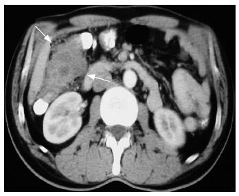
Figure 1 Computerized tomography revealed a 4 cm x 7 cm intraluminal mass originating from the ascending colon, adjacent to the right kidney and the liver, infiltrating the pericolic fat.
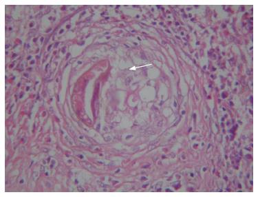
Figure 2 Histological study shows a trapped egg (arrow) in the center of a granuloma.
Eosinophilia is striking around this structure (HE, × 400).
- Citation: Makay O, Gurcu B, Caliskan C, Nart D, Tuncyurek M, Korkut M. Ectopic fascioliasis mimicking a colon tumor. World J Gastroenterol 2007; 13(18): 2633-2635
- URL: https://www.wjgnet.com/1007-9327/full/v13/i18/2633.htm
- DOI: https://dx.doi.org/10.3748/wjg.v13.i18.2633


