Copyright
©2007 Baishideng Publishing Group Inc.
World J Gastroenterol. May 14, 2007; 13(18): 2629-2632
Published online May 14, 2007. doi: 10.3748/wjg.v13.i18.2629
Published online May 14, 2007. doi: 10.3748/wjg.v13.i18.2629
Figure 1 Photos of patients's tumour.
Pigmented lesions (café–au-lait spots) and neurofibromas on the anterior abdominal wall (A) and on the back region (B).
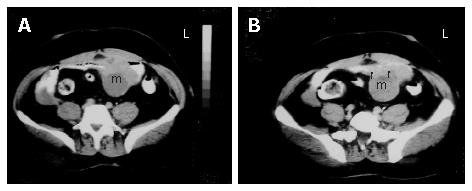
Figure 2 A: Axial sequential contrast-enhanced CT of the abdomen showing a well-defined heterogeneous mass consisting of an external solid area and internal cystic componenet adjacent to the anterior abdominal wall; B: the solid portions of the tumor were intensely stained on contrast imaging (arrows).
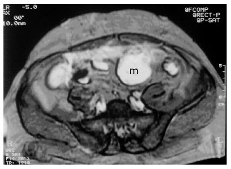
Figure 3 T2-weighted MRI shows heterogeneous hyperintense cystic and solid mass adjacent to the anterior abdominal wall.
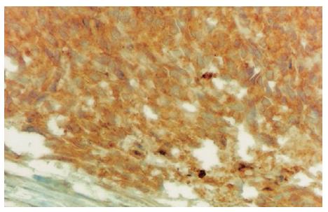
Figure 4 Immunohisto-chemical CD117 reactivity in tumor cells (DAB, × 400).
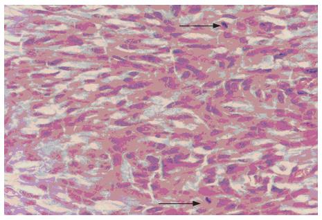
Figure 5 Spindle tumor cells with two mitoses marked with arrow (HE, × 400).
- Citation: Kalender ME, Sevinc A, Tutar E, Sirikci A, Camci C. Effect of sunitinib on metastatic gastrointestinal stromal tumor in patients with neurofibromatosis type 1: A case report. World J Gastroenterol 2007; 13(18): 2629-2632
- URL: https://www.wjgnet.com/1007-9327/full/v13/i18/2629.htm
- DOI: https://dx.doi.org/10.3748/wjg.v13.i18.2629


