Copyright
©2007 Baishideng Publishing Group Inc.
World J Gastroenterol. May 14, 2007; 13(18): 2535-2540
Published online May 14, 2007. doi: 10.3748/wjg.v13.i18.2535
Published online May 14, 2007. doi: 10.3748/wjg.v13.i18.2535
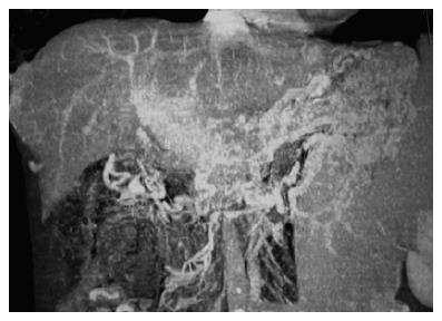
Figure 1 A case of chronic PVT in which typical cavernous transformation is observed.
The computed tomography angiography of the portal vein shows intensive collaterals around portal hilus and midline abdominal structures. Portal vein is not visible and replaced by collaterals.
- Citation: Harmanci O, Bayraktar Y. Portal hypertension due to portal venous thrombosis: Etiology, clinical outcomes. World J Gastroenterol 2007; 13(18): 2535-2540
- URL: https://www.wjgnet.com/1007-9327/full/v13/i18/2535.htm
- DOI: https://dx.doi.org/10.3748/wjg.v13.i18.2535


