Copyright
©2007 Baishideng Publishing Group Co.
World J Gastroenterol. Apr 21, 2007; 13(15): 2263-2265
Published online Apr 21, 2007. doi: 10.3748/wjg.v13.i15.2263
Published online Apr 21, 2007. doi: 10.3748/wjg.v13.i15.2263
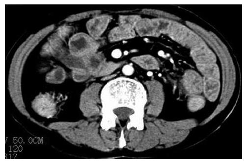
Figure 1 Abdominal computer tomography (CT) showing thickening of jejunal mucosa.
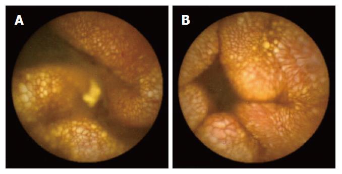
Figure 2 Capsule endoscopy showing diffuse oedematous aspect, dilatation of lymphatic vessels in the intestine (A) and thickening of whitish intestinal villi (B).
The dilated lymphatic vessels presented as coral and were mixed with dilated capillaries.
- Citation: Fang YH, Zhang BL, Wu JG, Chen CX. A primary intestinal lymphangiectasia patient diagnosed by capsule endoscopy and confirmed at surgery: A case report. World J Gastroenterol 2007; 13(15): 2263-2265
- URL: https://www.wjgnet.com/1007-9327/full/v13/i15/2263.htm
- DOI: https://dx.doi.org/10.3748/wjg.v13.i15.2263


