Copyright
©2007 Baishideng Publishing Group Co.
World J Gastroenterol. Apr 21, 2007; 13(15): 2258-2260
Published online Apr 21, 2007. doi: 10.3748/wjg.v13.i15.2258
Published online Apr 21, 2007. doi: 10.3748/wjg.v13.i15.2258
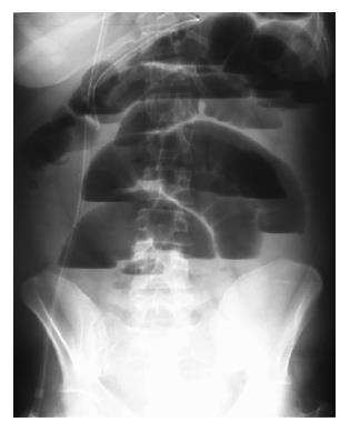
Figure 1 Abdominal X-ray revealing marked small bowel air-fluid levels.
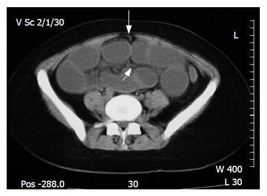
Figure 2 CT scan demonstrating dilated small bowel and a band originating from the umbilicus (big white arrow) and continuing between the small bowel loops (small white arrow).
- Citation: Markogiannakis H, Theodorou D, Toutouzas KG, Drimousis P, Panoussopoulos SG, Katsaragakis S. Persistent omphalomesenteric duct causing small bowel obstruction in an adult. World J Gastroenterol 2007; 13(15): 2258-2260
- URL: https://www.wjgnet.com/1007-9327/full/v13/i15/2258.htm
- DOI: https://dx.doi.org/10.3748/wjg.v13.i15.2258


