Copyright
©2007 Baishideng Publishing Group Co.
World J Gastroenterol. Apr 21, 2007; 13(15): 2255-2257
Published online Apr 21, 2007. doi: 10.3748/wjg.v13.i15.2255
Published online Apr 21, 2007. doi: 10.3748/wjg.v13.i15.2255
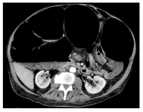
Figure 1 CT-scan showing massive distension of the colon (but not of the small intestine).
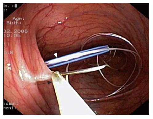
Figure 2 Endoscopic view of the left colon.
A 19-gauge needle grasped with an endoscopic snare (arrow) serves to hold the colon close to the abdominal wall while a string is passed through a Seldinger needle (arrowhead) to allow for transanal insertion of the colostomy tube.
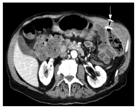
Figure 3 CT-scan showing the colostomy tube in place (arrow) and a normal-appearing colon.
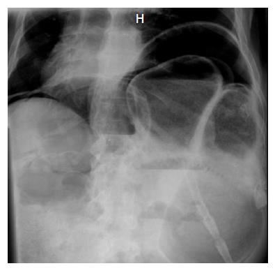
Figure 4 Abdominal plain X-ray showing a massive pneumoperitoneum.
- Citation: Bertolini D, De Saussure P, Chilcott M, Girardin M, Dumonceau JM. Severe delayed complication after percutaneous endoscopic colostomy for chronic intestinal pseudo-obstruction: A case report and review of the literature. World J Gastroenterol 2007; 13(15): 2255-2257
- URL: https://www.wjgnet.com/1007-9327/full/v13/i15/2255.htm
- DOI: https://dx.doi.org/10.3748/wjg.v13.i15.2255


