Copyright
©2007 Baishideng Publishing Group Co.
World J Gastroenterol. Apr 21, 2007; 13(15): 2250-2254
Published online Apr 21, 2007. doi: 10.3748/wjg.v13.i15.2250
Published online Apr 21, 2007. doi: 10.3748/wjg.v13.i15.2250
Figure 1 A: Chest CT before treatment.
A round and solid tumor seen in the right breast (white arrowheads). A solid and massive tumor shown in the lower mediastinum (white arrows) and the esophagus shifted to the left-front by the tumor (black arrowhead); B: Abdominal CT before treatment. Abdominal lymphadenopathy showing metastasis (black arrowheads); C: Esophagogram before treatment. Ulceration seen in the lower esophagus (white arrowhead), but there is no obstruction or stenosis.
Figure 2 A: The tumor consists of cells with little cytoplasm and round nuclei; HE (hematoxylin-eosin) stain (× 200); B: The tumor tissue diffusely expresses NSE; NSE (neuron-specific enolase) stain (× 200); C: The tumor tissue shows weakly positive expression; Keratin-wide stain (× 200).
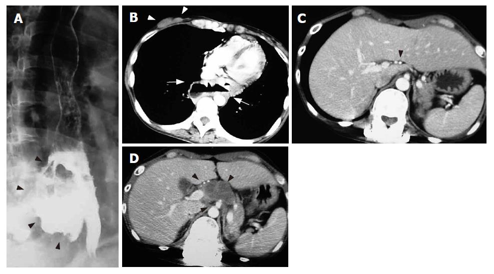
Figure 3 A: Esophagogram at 3 mo after initial treatment.
A giant esophagomediastinal fistula formed at the lower esophagus (black arrowheads); B: Three months after initial treatment. The massive mediastinal tumor completely disappeared with chemoradiotherapy, but a false lumen is shown at the same site (white arrows). No definite infection is observed. The right breast tumor also completely disappeared with chemoradiotherapy (white arrowheads); C: Three months after initial treatment. Abdominal lymphadenopathy completely disappeared with chemotherapy (black arrowhead); D: Two months after suspension of treatment (5 mo after initial treatment). Regrowth of abdominal tumor causing cancerous pain is shown (black arrowheads).
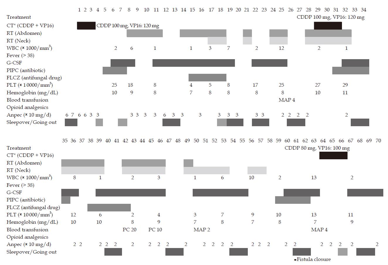
Figure 4 Course of treatment after restarting treatment (d 1) to closure of the fistula (d 60).
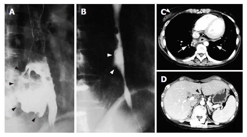
Figure 5 A: Esophagogram before restarting treatment (3 mo after initial treatment); B: Esophagogram 2 mo after restarting treatment (7 mo after the initial treatment).
The giant fistula was almost restored with good passage (white arrowheads); C: Chest CT 2 mo after restarting treatment. The CT reveals the esophagus repaired with normal mucosa but no recurrent tumor, and the false lumen forming a scar (white arrows). No recurrence is seen in the right breast (white arrowhead); D: The regrown abdominal tumor disappeared again (black arrowhead).
- Citation: Nomiya T, Teruyama K, Wada H, Nemoto K. Chemoradiotherapy for a patient with a giant esophageal fistula. World J Gastroenterol 2007; 13(15): 2250-2254
- URL: https://www.wjgnet.com/1007-9327/full/v13/i15/2250.htm
- DOI: https://dx.doi.org/10.3748/wjg.v13.i15.2250


