Copyright
©2007 Baishideng Publishing Group Co.
World J Gastroenterol. Mar 28, 2007; 13(12): 1870-1874
Published online Mar 28, 2007. doi: 10.3748/wjg.v13.i12.1870
Published online Mar 28, 2007. doi: 10.3748/wjg.v13.i12.1870
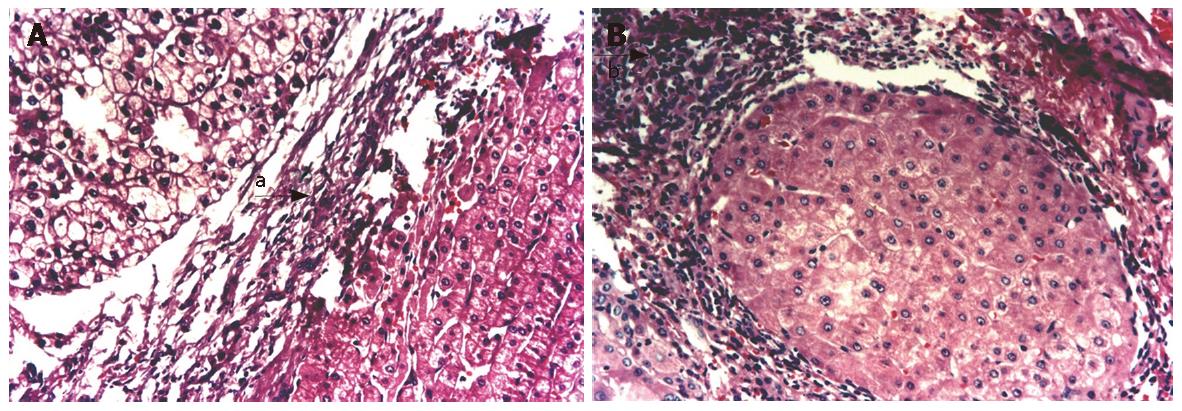
Figure 1 A: Fibrous connective tissues increased in surrounding HCC tissues, hepatic sclerotic tissue can be seen (arrow a); B: Inflammatory cells infiltrated surrounding noncancerous tissues (arrow b) (HE, × 200).
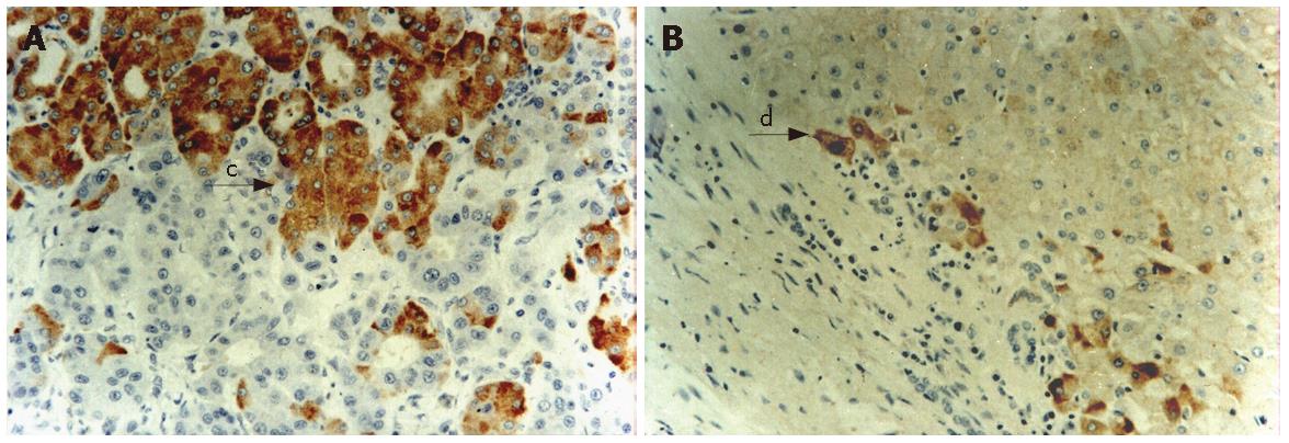
Figure 2 A: Expressions of HBsAg in HCC tissues (arrow c); B: Expressions of HCV in noncancerous tissues (arrow d) (Immunohistochemistry, SP, × 200).
- Citation: Xuan SY, Xin YN, Chen H, Shi GJ, Guan HS, Li Y. Significance of hepatitis B virus surface antigen, hepatitis C virus expression in hepatocellular carcinoma and pericarcinomatous tissues. World J Gastroenterol 2007; 13(12): 1870-1874
- URL: https://www.wjgnet.com/1007-9327/full/v13/i12/1870.htm
- DOI: https://dx.doi.org/10.3748/wjg.v13.i12.1870


