Copyright
©2007 Baishideng Publishing Group Co.
World J Gastroenterol. Mar 21, 2007; 13(11): 1755-1757
Published online Mar 21, 2007. doi: 10.3748/wjg.v13.i11.1755
Published online Mar 21, 2007. doi: 10.3748/wjg.v13.i11.1755
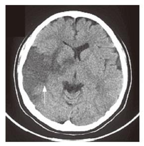
Figure 1 Plain computed tomography of brain indicates a low-density area in the right temporal lobe.
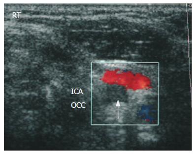
Figure 2 Cervical ultrasonography reveals a complete occlusion at the right internal carotid artery (arrow).
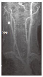
Figure 3 MRA shows occlusion at the right common carotid artery (arrow).
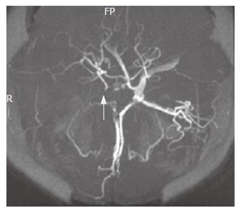
Figure 4 The right middle cerebral artery was not detectable (arrow).
- Citation: Nogami H, Iiai T, Maruyama S, Tani T, Hatakeyama K. Common carotid arterial thrombosis associated with ulcerative colitis. World J Gastroenterol 2007; 13(11): 1755-1757
- URL: https://www.wjgnet.com/1007-9327/full/v13/i11/1755.htm
- DOI: https://dx.doi.org/10.3748/wjg.v13.i11.1755


