Copyright
©2007 Baishideng Publishing Group Co.
World J Gastroenterol. Mar 21, 2007; 13(11): 1747-1751
Published online Mar 21, 2007. doi: 10.3748/wjg.v13.i11.1747
Published online Mar 21, 2007. doi: 10.3748/wjg.v13.i11.1747
Figure 1 IL-12 and IL-18 expression in gastric cancer tissues (× 400).
A: IL-12 expression appears predominantly in the cytoplasm of gastric cancer cells; B: IL-18 is present in both the cytoplasm and nuclei of gastric cancer cells.
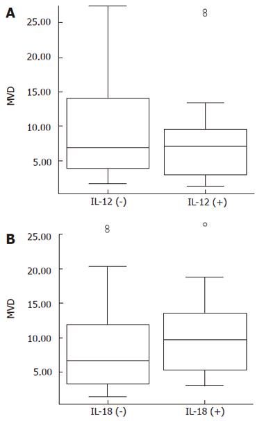
Figure 2 The association between MVD and (A) IL-12, (B) IL-18.
Boxes correspond to interquartile ranges. Lines in boxes represent the median values. The open circles represent the outliers.
Figure 3 Kaplan-Meier survival curves of patients with gastric cancer.
A: IL-12-positive tumor has a significantly lower overall survival than those with IL-12-negative tumor (P = 0.002); B: The positive or negative expression of IL-18 shows no difference in overall survival (P = 0.342).
- Citation: Ye ZB, Ma T, Li H, Jin XL, Xu HM. Expression and significance of intratumoral interleukin-12 and interleukin-18 in human gastric carcinoma. World J Gastroenterol 2007; 13(11): 1747-1751
- URL: https://www.wjgnet.com/1007-9327/full/v13/i11/1747.htm
- DOI: https://dx.doi.org/10.3748/wjg.v13.i11.1747


