Copyright
©2007 Baishideng Publishing Group Co.
World J Gastroenterol. Mar 21, 2007; 13(11): 1696-1700
Published online Mar 21, 2007. doi: 10.3748/wjg.v13.i11.1696
Published online Mar 21, 2007. doi: 10.3748/wjg.v13.i11.1696
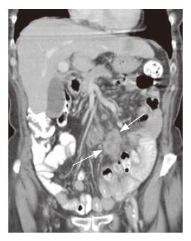
Figure 1 Cor-onal reconstruc-tion in a patient with RCD typeIIshowing cavitation of lymphnodes and smaller non cavitated lymphnodes in combination with infiltrated mesenterial fat (arrows).
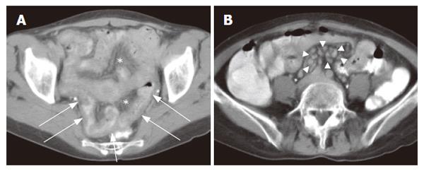
Figure 2 Axial CT image of a patient with RCD typeIdemonstrates (A) multiple small lymph nodes (arrowheads) and (B) thickening of the ileal wall (arrows) and infiltration of the mesenteric fat (*).
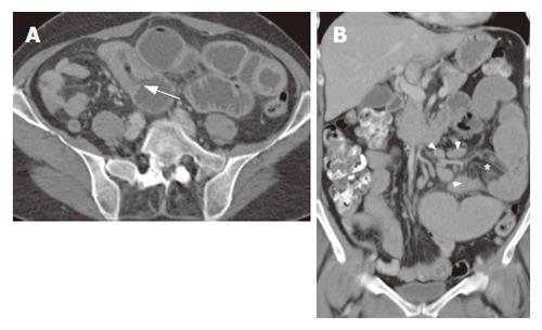
Figure 3 Axial CT image (A) demonstrates EATL (arrow) and prestenotic dilatation of jejunal loops.
The coronal reformation (B) demonstrates dilated jejunum with infiltrated mesenterial fat (*) and multiple enlarged lymph nodes (arrowheads).
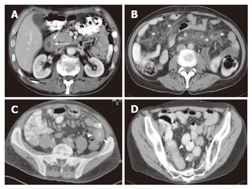
Figure 4 Axial CT images demonstrating locations of EATL.
A: Duodenal localization of lymphoma (arrow); B: Mesenterial localization showing “misty mesenterium” (*) and multiple lymphnodes; C: Jejunal localization of lymphoma (arrowhead) and multiple lymphnodes; D: Ileal localization (arrowhead).
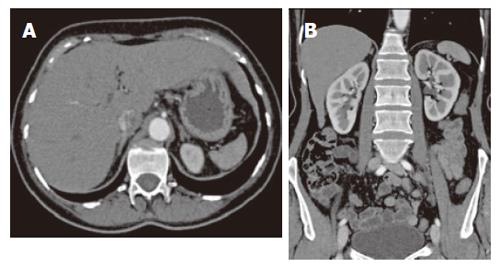
Figure 5 Hyposplenism in a patient with RCD typeI demonstrated on axial image (A) and coronal (B) reconstruction.
- Citation: Mallant M, Hadithi M, Al-Toma AB, Kater M, Jacobs M, Manoliu R, Mulder C, van Waesberghe JH. Abdominal computed tomography in refractory coeliac disease and enteropathy associated T-cell lymphoma. World J Gastroenterol 2007; 13(11): 1696-1700
- URL: https://www.wjgnet.com/1007-9327/full/v13/i11/1696.htm
- DOI: https://dx.doi.org/10.3748/wjg.v13.i11.1696


