Copyright
©2007 Baishideng Publishing Group Co.
World J Gastroenterol. Mar 21, 2007; 13(11): 1659-1665
Published online Mar 21, 2007. doi: 10.3748/wjg.v13.i11.1659
Published online Mar 21, 2007. doi: 10.3748/wjg.v13.i11.1659
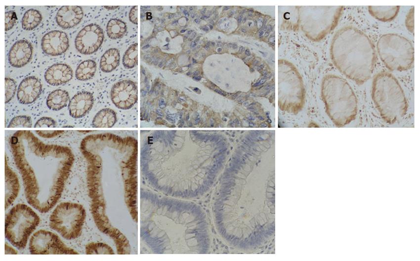
Figure 1 Expression of cytokeratin AE1/AE3 and PCNA in primary colorectal adenocarcinomas.
We performed immunohistochemistry with cytokeratin AE1/AE3 antibodies showing positive staining in cytoplasm of colorectal normal crypts (A) and malignant crypts (B) (positive in brown). While the proliferation marker PCNA stained the nuclei of the normal (C) and tumor (D) tissues (positive in brown). E is the negative control.
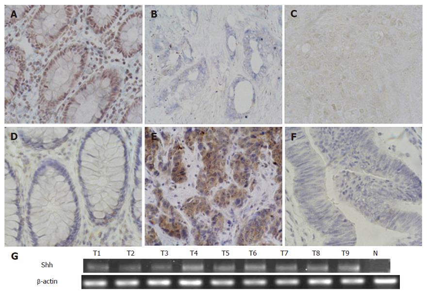
Figure 2 Expression of Shh in primary colorectal adenocarcinomas.
In situ hybridization was performed to detect Shh transcript in normal (A) and cancerous (B) tissues (positive in blue), and the sense probe did not reveal any positive signals (C is the sense control of B). Results of in situ hybridization were confirmed by immunohistostaining in normal (D) and tumor (E) tissues (positive in brown, F is the negative control) and by RT-PCR (G).
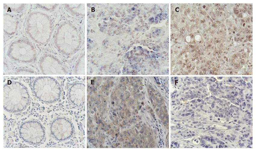
Figure 3 Expression of Ptch1 in primary colorectal adenocarcinomas.
Ptch1 transcript (blue as positive) was detected by in situ hybridization in the normal control (A) and colorectal cancers (B). C is from the same tumor with B being derived from the Ptch1 sense probe. To confirm the in situ hybridization results, we performed immunohistochemistry with PTCH1 antibodies, showing positive staining of PTCH1 protein (positive in brown) in malignant crypts (E). D is the normal control and F is the negative control.
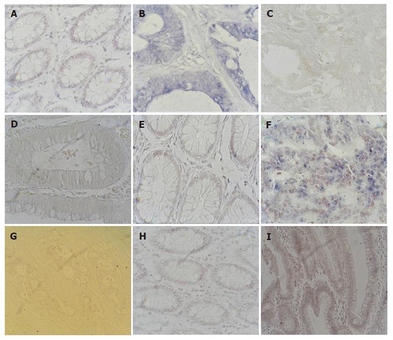
Figure 4 Expression of Gli1, Gli3 and Hip in primary colorectal adenocarcin-omas.
We detected expr-ession patterns of Gli1, Gli3 and Hip transcripts by in situ hybridization (positive in blue). Gli1 (A) and Hip (E) were not expressed in normal control tissue. B: Gli1 was expressed in cytop-lasm of malignant crypt. The expression of Gli1 (C) and Hip (D) sense probe in tumor tissue. F: Hip was expressed in cytoplasm of tumor cells. G: Gli3 was not expressed in colorectal adenocarcinomas. H is the normal control of Gli3 and I is the sense control of Gli3.
Figure 5 Expression of PDGFRα in primary colorectal adenocarcinomas.
A: We analyzed expression pattern of PDGFRα transcript by in situ hybridization (blueas positive); B: Expression of PDGFRα in normal control was high in aberrant crypts and moderate in the stroma; C: The expression of PDGFR sense probe in tumor tissue.
- Citation: Bian YH, Huang SH, Yang L, Ma XL, Xie JW, Zhang HW. Sonic hedgehog-Gli1 pathway in colorectal adenocarcinomas. World J Gastroenterol 2007; 13(11): 1659-1665
- URL: https://www.wjgnet.com/1007-9327/full/v13/i11/1659.htm
- DOI: https://dx.doi.org/10.3748/wjg.v13.i11.1659


