Copyright
©2007 Baishideng Publishing Group Co.
World J Gastroenterol. Mar 21, 2007; 13(11): 1652-1658
Published online Mar 21, 2007. doi: 10.3748/wjg.v13.i11.1652
Published online Mar 21, 2007. doi: 10.3748/wjg.v13.i11.1652
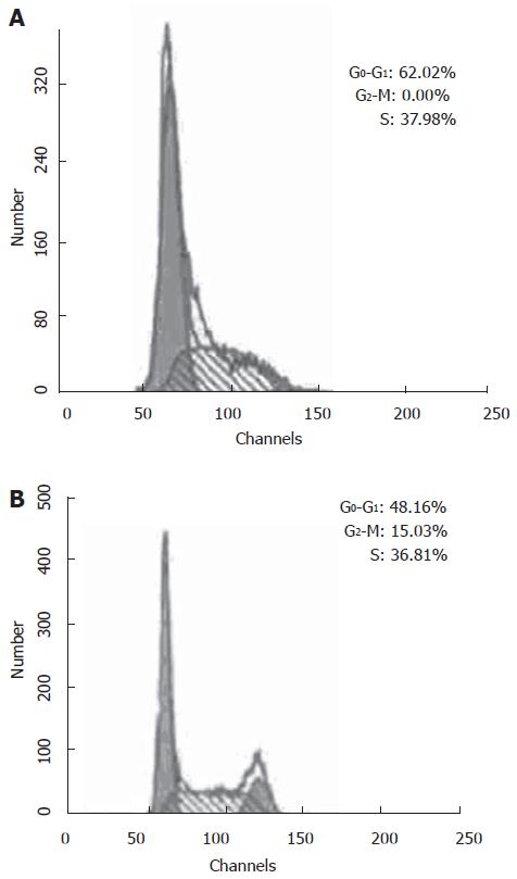
Figure 1 A: Cell cycle distribution of BEL7402 treated with UDCA (0.
8 mmol/L) for 48 h. The cell cycle distribution changed, the proportion of G0-G1 phase increased significantly (P < 0.05) and the proportion of S phase and G2-M phase decreased significantly (P < 0.05); B: Cell cycle distribution of L-02 cells treated with UDCA (0.8 mmol/L) for 48 h. The cell cycle distribution of L-02 cells does not change.
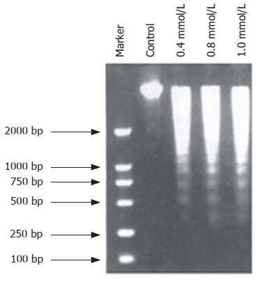
Figure 2 Induction of internucleosomal DNA fragmentation by UDCA.
Cells were incubated without or with various concentrations of UDCA (0.4 mmol/L, 0.8 mmol/L, 1.0 mmol/L) for 48 h. DNA was extracted and analyzed by 1.5% agarose gel electrophoresis in the presence of ethidium bromide staining.
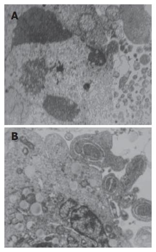
Figure 3 Morphology of HepG2 treated with UDCA (0.
4 mmol/L) for 48 h (TEM, × 4000). A: Nuclear frag-mentation,chromosome condensation, cell shrinkage and loss of cell-cell contact are visible; B: Subsequent ruffling and blebbling of the cell membrane, and formation of apoptotic bodies are also observed.
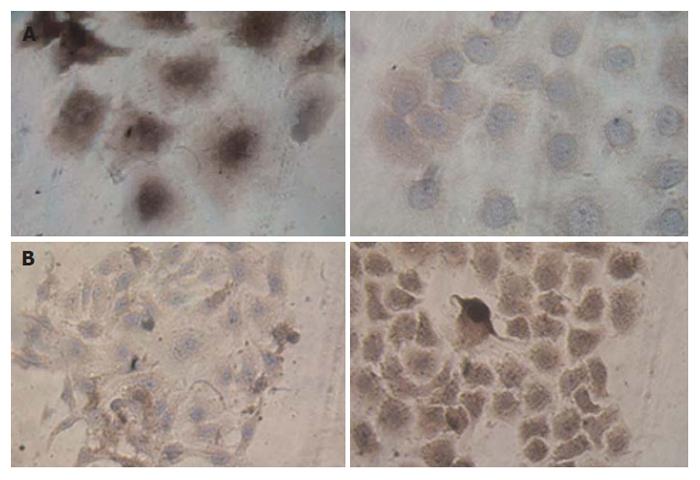
Figure 4 A: Effect of UDCA on expression of bcl-2 in BEL7402, immuno-cytochemical staining of bcl-2.
The accumulation of bcl-2 protein is indicated by a brown coloration in the cytoplasm (× 200). Control group, expression of bcl-2 in BEL7402 cultivated for 48 h (Left), Expression of bcl-2 in BEL7402 treated with UDCA (0.4 mmol/L) for 48 h (Right); B: Effect of UDCA on expression of Bax in BEL7402, immunocytochemical staining of Bax. The accumulation of Bax protein is indicated by a brown coloration in the cytoplasm. Control group,expression of Bax in BEL7402 cultivated for 48 h (Left). Expression of Bax in BEL7402 treated with UDCA (0.4 mmol/L) 48 h (× 200) (Right).
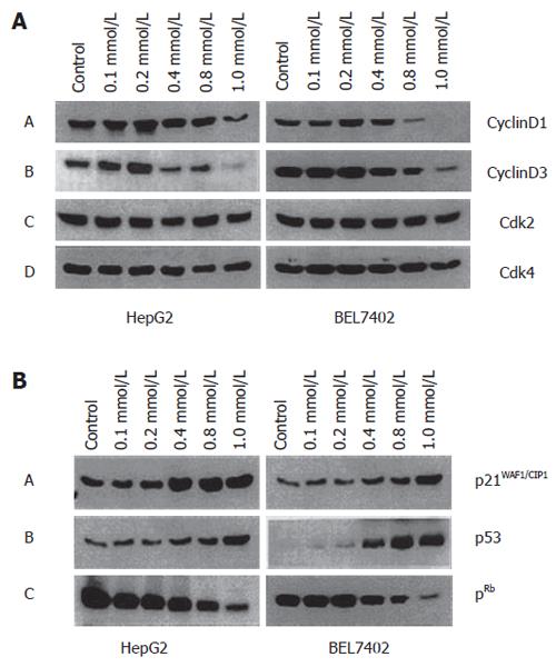
Figure 5 A: Effects of UDCA on production of cyclins and Cdks in HepG2 and BEL7402 cells treated with various concentrations of UDCA (0.
1, 0.2, 0.4, 0.8, 1.0 mmol/L) for 48 h; B: Effects of UDCA treatment on protein levels of p21WAF1/CIP1, p53, pRb in HepG2 and BEL7402. The cells were treated with 0.8 mmol/L UDCA for 48 h.
- Citation: Liu H, Qin CY, Han GQ, Xu HW, Meng M, Yang Z. Mechanism of apoptotic effects induced selectively by ursodeoxycholic acid on human hepatoma cell lines. World J Gastroenterol 2007; 13(11): 1652-1658
- URL: https://www.wjgnet.com/1007-9327/full/v13/i11/1652.htm
- DOI: https://dx.doi.org/10.3748/wjg.v13.i11.1652


