Copyright
©2006 Baishideng Publishing Group Co.
World J Gastroenterol. Mar 7, 2006; 12(9): 1386-1391
Published online Mar 7, 2006. doi: 10.3748/wjg.v12.i9.1386
Published online Mar 7, 2006. doi: 10.3748/wjg.v12.i9.1386
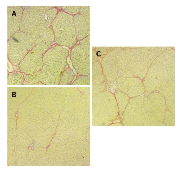
Figure 1 Effects of IL-10 on histology of CC4-induced fibrotic rat liver after treated with CCL4 for 9 wk (A), IL-10 for 3 wk (B) and spontaneous recovery for 3 wk (C).
Figure 2 Photomicrographs of liver tissue from rats after treatment with with CCL4 (A), IL-10 for 3 wk (B) and spontaneous recovery for 3 wk(C) by Picrosirius staining (40x).
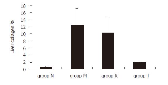
Figure 3 Percentage of collagen types I and III in fibrotic rats.
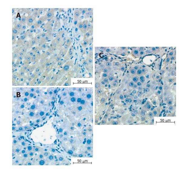
Figure 4 Expression of MMP-2 protein in liver tissue of rats treated with CCL4 for 9 wk (A), IL-10 for 3 wk (B) and spontaneous recovery for 3 wk (C).
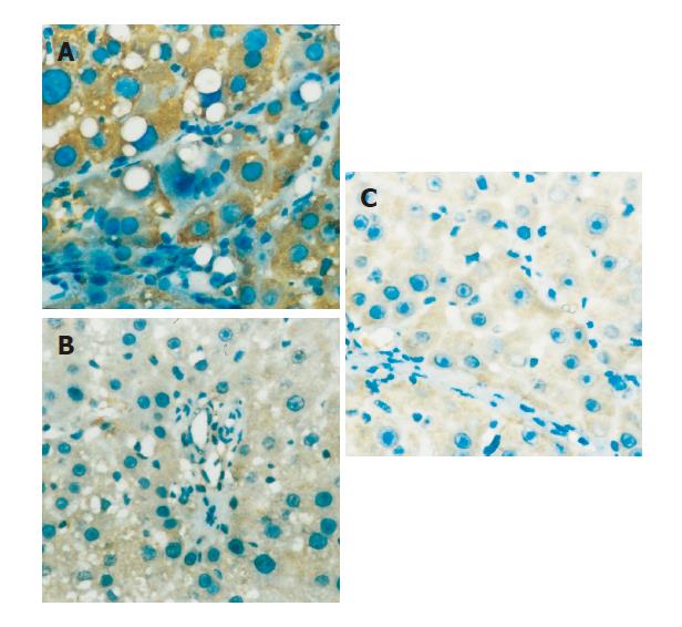
Figure 5 Expression of TIMP-1 protein in liver tissue of rats treated with CCL4 for 9 wk (A), IL-10 for 3 wk (B) and spontaneous recovery for 3 wk (C).
Figure 6 Relative expression levels of MMP-2 and TIMP-1 in liver of different groups.
Figure 7 Expression of TNF-a protein in liver tissue of rats treated with CCL4 for 9 wk (A), IL-10 for 3 wk (B) and spontaneous recovery for 3 wk (C).
Figure 8 Relative expression of TNF-α in rat liver of different groups.
- Citation: Huang YH, Shi MN, Zheng WD, Zhang LJ, Chen ZX, Wang XZ. Therapeutic effect of interleukin-10 on CCl4-induced hepatic fibrosis in rats. World J Gastroenterol 2006; 12(9): 1386-1391
- URL: https://www.wjgnet.com/1007-9327/full/v12/i9/1386.htm
- DOI: https://dx.doi.org/10.3748/wjg.v12.i9.1386


