Copyright
©2006 Baishideng Publishing Group Co.
World J Gastroenterol. Feb 14, 2006; 12(6): 982-986
Published online Feb 14, 2006. doi: 10.3748/wjg.v12.i6.982
Published online Feb 14, 2006. doi: 10.3748/wjg.v12.i6.982
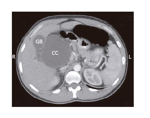
Figure 1 Upper abdominal CT (after intravenous administration of contrast material) demonstrating the extrahepatic portion of the choledochal cyst (CC).
GB: gallbladder.
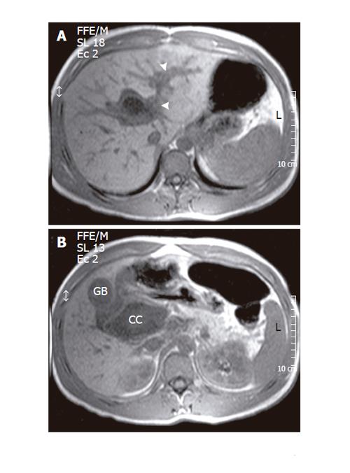
Figure 2 Significant distension of the intrahepatic bile ducts (arrowhead) in MRI of the upper abdomen (gradient echo T1-weighed acquisition) (A).
The walls of the choledochal cyst (CC) tend to acquire a concave shape, while its volume appears reduced as compared to the CT findings, both signs are considered as indicative of rupture (B). GB: gallbladder.
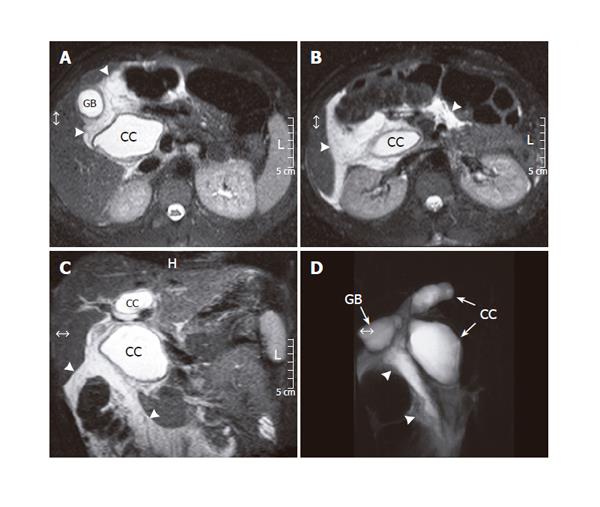
Figure 3 Abdominal MRI (spin echo T2-weighed, transverse projections - A and B, coronal projection - C) displaying bile leakage into the peritoneal cavity (arrowheads).
The study was followed by MRCP (turbo spin echo T2-weighed acquisition - D) which imaged the bile leak (arrowheads) and moreover revealed the “egg-timer” bicameral shape of the extrahepatic moiety of the choledochal cyst (CC). GB: gallbladder.
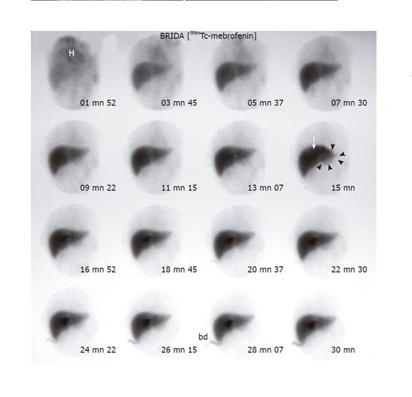
Figure 4 Postoperative 99mTc-mebrofenin dynamic study.
A well-circumscribed fusiform tracer accumulation in the right hepatic lobe near the hepatic hilum is clearly visible from the 15th min of the study (arrow), and corresponds to a dilated intrahepatic duct. A much fainter bulbous accumulation is apparent in the area of the left hepatic lobe, extending well beyond the liver margins (arrowheads) and corresponding to the extrahepatic portion of the choledochal cyst. H: heart blood pool; bd: biliary drainage.
Figure 5 Planar static scintigraphic images of the upper abdomen.
At 90-min post-injection the faint tracer accumulation corresponding to the extrahepatic portion of the choledochal cyst (CC) extends beyond the liver margins (arrowheads), while a fusiform tracer accumulation in a cystic dilated intrahepatic duct is prominent (arrow) (A). The latter finding is not visible in the late (24 h) image, nor bowel or peritoneal activity (B). bd: biliary drainage; K: kidney; S: spleen.
- Citation: Stipsanelli E, Valsamaki P, Tsiouris S, Arka A, Papathanasiou G, Ptohis N, Lahanis S, Papantoniou V, Zerva C. Spontaneous rupture of a type IVA choledochal cyst in a young adult during radiological imaging. World J Gastroenterol 2006; 12(6): 982-986
- URL: https://www.wjgnet.com/1007-9327/full/v12/i6/982.htm
- DOI: https://dx.doi.org/10.3748/wjg.v12.i6.982


