Copyright
©2006 Baishideng Publishing Group Co.
World J Gastroenterol. Feb 14, 2006; 12(6): 971-973
Published online Feb 14, 2006. doi: 10.3748/wjg.v12.i6.971
Published online Feb 14, 2006. doi: 10.3748/wjg.v12.i6.971
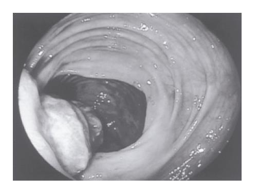
Figure 1 Endoscopic appearance of polypoidal lesion in the cecum.
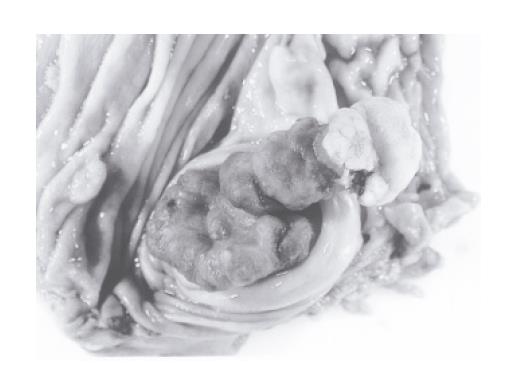
Figure 2 Gross specimen of invaginated appendiceal tumor seen on opening cecum.
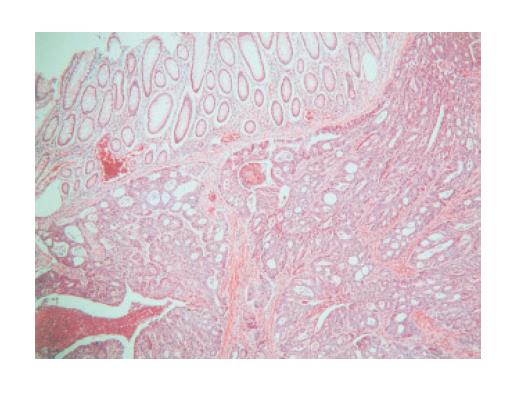
Figure 3 Well-differentiated neuroendocrine carcinoma, arranged in nests and trabeculae, beneath uninvolved large-intestinal type appendiceal mucosa (top left hand corner) (hematoxylin and eosin; ×16).
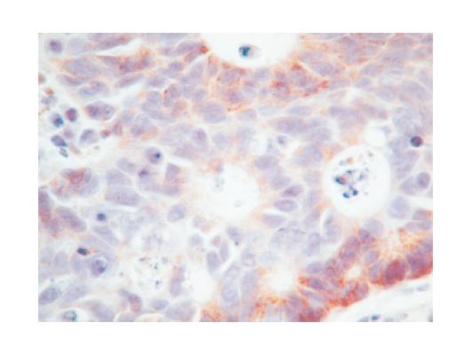
Figure 4 Chromogranin positive immunohistochemical staining (brown cytoplasmic staining) demonstrating neuroendocrine differentiation of tumor cells (×40, 1:5 000 dilution, DAK-A3 clone, DAKO, Cambridge, UK).
- Citation: Thomas RE, Maude K, Rotimi O. A case of an intussuscepted neuroendocrine carcinoma of the appendix. World J Gastroenterol 2006; 12(6): 971-973
- URL: https://www.wjgnet.com/1007-9327/full/v12/i6/971.htm
- DOI: https://dx.doi.org/10.3748/wjg.v12.i6.971


