Copyright
©2006 Baishideng Publishing Group Co.
World J Gastroenterol. Feb 14, 2006; 12(6): 921-927
Published online Feb 14, 2006. doi: 10.3748/wjg.v12.i6.921
Published online Feb 14, 2006. doi: 10.3748/wjg.v12.i6.921
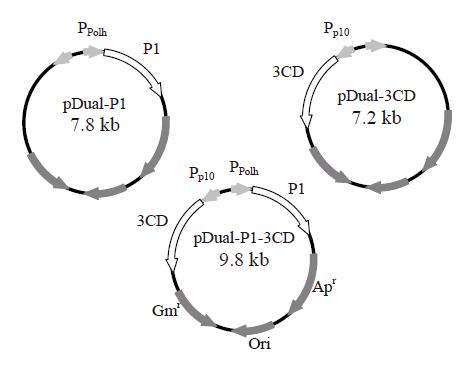
Figure 1 Construction of three recombinant baculovirus vectors using pFastBacTM DUAL plasmid as the backbone.
The gene fragments encoding P1 and 3CD were cloned separately into MCS I and II of pFastBacTM DUAL under the control of polyhedrin (Ppolh) and p10 promoters (Pp10), respectively. The resultant plasmids were designated pDual-P1 and pDual-3CD, respectively. The P1 and 3CD genes were cloned together into MCS I and II under the control of polyhedrin and p10 promoters. The resultant plasmid was designated pDual-P1-3CD.
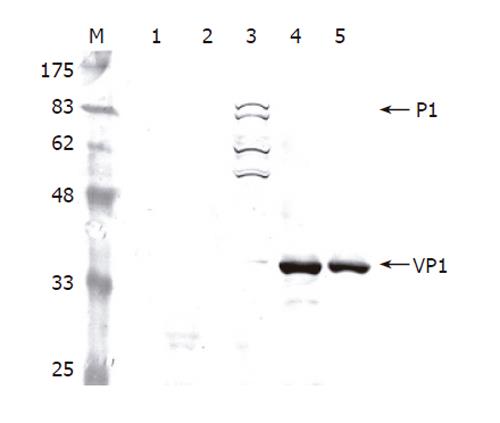
Figure 2 Western blot analysis of cell lysates infected by different viruses.
The Sf-9 cells were infected by the viruses at a total MOI of 10 and harvested at 3 dpi. The proteins were separated by SDS-PAGE, electrotransferred to a nitrocellulose membrane and probed using anti-VP1 MAb as the primary antibody. Lane 1: mock infection; Lane 2: wild-type baculovirus AcMNPV; Lane 3: Bac-P1; Lane 4: Bac-P1-3CD; Lane 5: Bac-P1 and Bac-3CD.
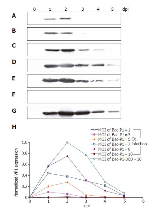
Figure 3 Time course profiles of VP1 production under various infection conditions as analyzed by Western blot.
Insect cells were infected at a total MOI of 10 by single infection with Bac-P1-3CD (G), or co-infection at MOI ratios (Bac-P1/Bac-3CD) of 1/9 (b), 3/7 (B), 5/5 (C), 7/3 (D), 9/1 (E) and 10/0 (F). Lanes 0-5 represent the cell lysates collected at 0, 1, 2, 3, 4, and 5 dpi, respectively. The relative VP1 production levels under different conditions were quantified by scanning densitometry (H). The data represent the average values of duplicated experiments.
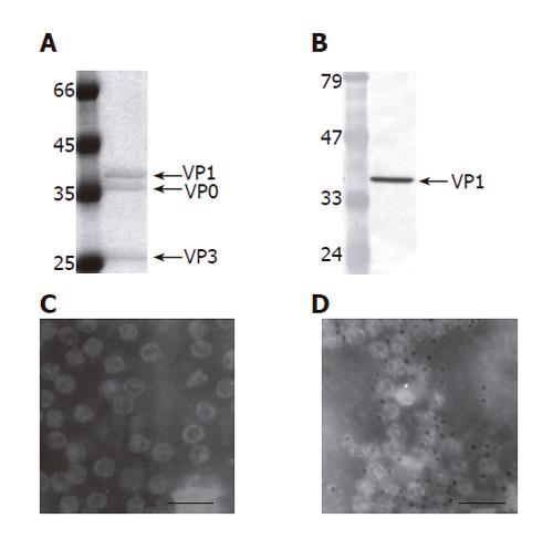
Figure 5 Purification and characterization of EV71 VLP (100 000 X magnification,).
The VLPs were produced by infecting 150 mL Sf-9 cells (1.5×10⁶ cells/mL) with Bac-P1-3CD at MOI 10, harvested at 2 dpi and purified by ultracentrifugation. The purified VLPs were characterized by SDS-PAGE (A), Western blot (B), TEM examination (C) and immunogold labeling (D). The PAGE gel was stained by Commassie blue and scanned for densitometry analysis. The primary antibody used in the Western blot and immunogold labeling was anti-VP1 MAb. The size of the gold particles was 5 nm. Bar: 100 nm.
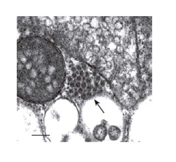
Figure 4 Electron microscopy examinations of EV71 VLP aggregates formed in cytoplasm of the Bac-P1-3CD-infected insect cells (100 000 X magnification).
The cells were infected at MOI 10, harvested at 3 dpi and ultrathin sectioned for TEM. The arrowhead indicates the VLP aggregate. Bar:100 nm.
- Citation: Chung YC, Huang JH, Lai CW, Sheng HC, Shih SR, Ho MS, Hu YC. Expression, purification and characterization of enterovirus-71 virus-like particles. World J Gastroenterol 2006; 12(6): 921-927
- URL: https://www.wjgnet.com/1007-9327/full/v12/i6/921.htm
- DOI: https://dx.doi.org/10.3748/wjg.v12.i6.921


