Copyright
©2006 Baishideng Publishing Group Co.
World J Gastroenterol. Dec 28, 2006; 12(48): 7859-7863
Published online Dec 28, 2006. doi: 10.3748/wjg.v12.i48.7859
Published online Dec 28, 2006. doi: 10.3748/wjg.v12.i48.7859
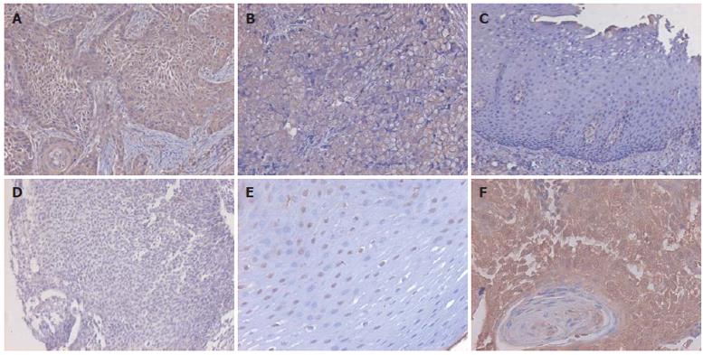
Figure 1 Immunohistochemical analysis of Elk-1 in paired ESCC samples using anti-Elk-1 antibody (1:100) showing diffuse and strong staining in cytoplasm of esophageal cancer epithelial cells well-differentiated tumor (A), moderately-differentiated tumor (B), sporadic and weak staining in the cytoplasm of normal epithelial cells (C), negative control designed using PBS instead of primary antibody (D), strong staining in nuclei of normal epithelial cells (E) (A-E × 100), and in cytoplasm of well-differentiated esophageal cancer epithelial cells (F) (× 200).
- Citation: Chen AG, Yu ZC, Yu XF, Cao WF, Ding F, Liu ZH. Overexpression of Ets-like protein 1 in human esophageal squamous cell carcinoma. World J Gastroenterol 2006; 12(48): 7859-7863
- URL: https://www.wjgnet.com/1007-9327/full/v12/i48/7859.htm
- DOI: https://dx.doi.org/10.3748/wjg.v12.i48.7859


