Copyright
©2006 Baishideng Publishing Group Co.
World J Gastroenterol. Dec 28, 2006; 12(48): 7774-7778
Published online Dec 28, 2006. doi: 10.3748/wjg.v12.i48.7774
Published online Dec 28, 2006. doi: 10.3748/wjg.v12.i48.7774
Figure 1 Hybrid HBcAg-T protein showing HCV T epitope (hatched box) inserted between HBcAg aa 79 and 80, a region located at the tip of core particle surface spikes.
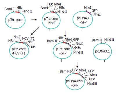
Figure 2 The construction of plasmids pTrc-core-HCV (T) and pcDNA3.
1-core-GFP.
Figure 3 Hybrid HBcAg-GFP protein showing the GFP protein (hatched box) inserted between HBcAg aa 79 and 80.
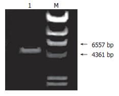
Figure 4 Plasmid pTrc-coreNheI identified by restriction endonuclease NheI cleavage.
Land 1: pTrc-coreNheI digest; M: λ-HindIII digest.
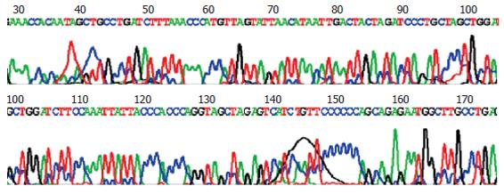
Figure 5 The sequencing of plasmid pTrc-coreNheI PCR product.
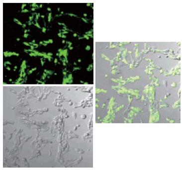
Figure 6 pcDNA3.
1-HBc-GFP expression in Hela cell line (confocal microscopy, 300 ×).
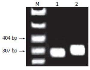
Figure 7 PCR products of pTrc-core-HCV(T).
Lanes 1-2: pTrc-coreNheI or pTrc-core-HCV (T) PCR product; M: pBR322 DNA-Msp I digest.
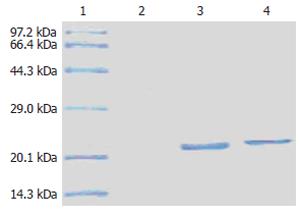
Figure 8 The results of HBcAg-T and HBcAg by SDS-PAGE.
1: Marker; 2: Negative; 3: HBcAg; 4: HBcAg-T.
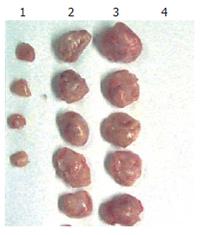
Figure 9 Tumor regression trial in mice.
1: HBcAg immunized team; 2,3: Vacuity team; 4: HBcAg-T immunized team.
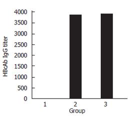
Figure 10 The titer of HBcAb.
1: Vacuity team; 2: HBcAg immunized team; 3:HBcAg-T immunized team.
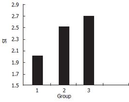
Figure 11 Nonspecific lymphocytes prolifera-tive response.
1: Blank team; 2: HBcAg immunized team; 3: HBcAg-T immunized team.
- Citation: Chen JY, Li F. Development of hepatitis C virus vaccine using hepatitis B core antigen as immuno-carrier. World J Gastroenterol 2006; 12(48): 7774-7778
- URL: https://www.wjgnet.com/1007-9327/full/v12/i48/7774.htm
- DOI: https://dx.doi.org/10.3748/wjg.v12.i48.7774


