Copyright
©2006 Baishideng Publishing Group Co.
World J Gastroenterol. Nov 28, 2006; 12(44): 7203-7205
Published online Nov 28, 2006. doi: 10.3748/wjg.v12.i44.7203
Published online Nov 28, 2006. doi: 10.3748/wjg.v12.i44.7203
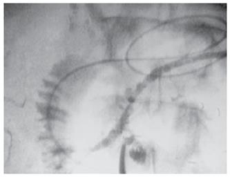
Figure 2 Intraoperative pancreatography using an ENPD tube demonstrates a communicating duct between the main pancreatic duct and cystic lesion.
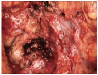
Figure 3 Operative photograph shows the pancreas after completion of the single-duct resection of the pancreas for multiple IPMNs.
Arrows indicate the cut surface after pancreatic resection.
- Citation: Kuroki T, Tajima Y, Tsutsumi R, Tsuneoka N, Kitasato A, Adachi T, Kanematsu T. Endoscopic naso-pancreatic stent-guided single-branch resection of the pancreas for multiple intraductal papillary mucinous adenomas. World J Gastroenterol 2006; 12(44): 7203-7205
- URL: https://www.wjgnet.com/1007-9327/full/v12/i44/7203.htm
- DOI: https://dx.doi.org/10.3748/wjg.v12.i44.7203


