Copyright
©2006 Baishideng Publishing Group Co.
World J Gastroenterol. Nov 21, 2006; 12(43): 7051-7054
Published online Nov 21, 2006. doi: 10.3748/wjg.v12.i43.7051
Published online Nov 21, 2006. doi: 10.3748/wjg.v12.i43.7051
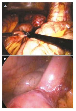
Figure 1 Laparoscopic laparotomy showing small intestinal bleeding (A and B).
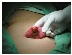
Figure 2 An oval stromal tumor causing small intestinal bleeding.
Laparoscopic laparotomy found there is an oval stromal tumor on the up segment of jejunum, with clear borderline and smooth surface, having no adhesion with other tissue around it. Expansive abdominal excision was performed to draw out the tumor for resection.
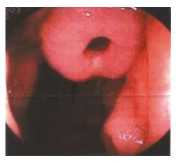
Figure 3 Electric intestinal endoscopy showing a benign jejunum tumor.
A deep ulcer sunken on the top of the tumor could be seen.
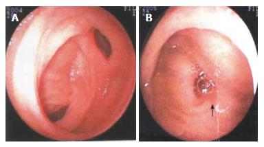
Figure 4 Double balloon enteroscopy showing the clamped diseased intestinal segment (A) and resected diseased intestinal tract (B).
The double-balloon enteroscope was pushed 200 cm into the ileum through anus. Diverticulum was found in the ileum 90-100 cm away from the ileocecal valve, at the opening of which a 1.2 cm × 1.0 cm ulcer was observed. The ulcer had thin covering of lichenoid substance, but no active bleeding. No other abnormalities were found. Meckel’s diverticulum was diagnosed.
- Citation: Ba MC, Qing SH, Huang XC, Wen Y, Li GX, Yu J. Application of laparoscopy in diagnosis and treatment of massive small intestinal bleeding: Report of 22 cases. World J Gastroenterol 2006; 12(43): 7051-7054
- URL: https://www.wjgnet.com/1007-9327/full/v12/i43/7051.htm
- DOI: https://dx.doi.org/10.3748/wjg.v12.i43.7051


