Copyright
©2006 Baishideng Publishing Group Co.
World J Gastroenterol. Nov 21, 2006; 12(43): 7042-7046
Published online Nov 21, 2006. doi: 10.3748/wjg.v12.i43.7042
Published online Nov 21, 2006. doi: 10.3748/wjg.v12.i43.7042
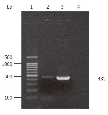
Figure 1 Electrophoresis of HP-NAP PCR products.
Lane 1: 100 bp DNA ladder marker; lanes 2 and 3: PCR products of HP-NAP; lane 4: Blank control.
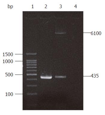
Figure 2 Identification map of recombinant plasmid pIRES-NAP.
Lane 1: 100 bp DNA ladder marker; lane 2: PCR products templated on pIRES-NAP; lane 3: pIRES-NAP digested by endonucleases Xho I and Mlu I; lane 4: Blank control.
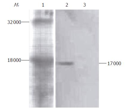
Figure 3 Western blot analysis of HP-NAP fusion protein expression.
Lane 1: Protein standard; lane 2: COS-7 cells transfected with pIRES-NAP; lane 3: COS-7 cells without transfection as control.
-
Citation: Sun B, Li ZS, Tu ZX, Xu GM, Du YQ. Construction of an oral recombinant DNA vaccine from
H pylori neutrophil activating protein and its immunogenicity. World J Gastroenterol 2006; 12(43): 7042-7046 - URL: https://www.wjgnet.com/1007-9327/full/v12/i43/7042.htm
- DOI: https://dx.doi.org/10.3748/wjg.v12.i43.7042


