Copyright
©2006 Baishideng Publishing Group Co.
World J Gastroenterol. Nov 14, 2006; 12(42): 6889-6892
Published online Nov 14, 2006. doi: 10.3748/wjg.v12.i42.6889
Published online Nov 14, 2006. doi: 10.3748/wjg.v12.i42.6889
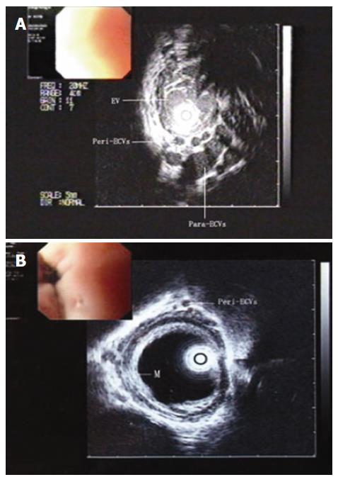
Figure 1 Alteration in appearance of ultrasound microprobe images.
A: Before treatment, enlarged tortuous varices were found inside of the esophageal wall, while many small vessels adjacent to the muscularis externa formed a venous plexus. B: After treatment, anechoic areas inside and outside of the esophageal wall disappeared or remained only tiny, while the echo of mucosa and submucosa of the esophagus enhanced. EV: Varices inside of the esophageal wall; Peri-ECVs: periesophageal collateral veins; Para-ECVs: Paraesophageal collateral veins; M: Esophageal mucosa.
- Citation: Liu B, Deng MH, Lin N, Pan WD, Ling YB, Xu RY. Evaluation of the effects of combined endoscopic variceal ligation and splenectomy with pericardial devascularization on esophageal varices. World J Gastroenterol 2006; 12(42): 6889-6892
- URL: https://www.wjgnet.com/1007-9327/full/v12/i42/6889.htm
- DOI: https://dx.doi.org/10.3748/wjg.v12.i42.6889


