Copyright
©2006 Baishideng Publishing Group Co.
World J Gastroenterol. Nov 7, 2006; 12(41): 6646-6651
Published online Nov 7, 2006. doi: 10.3748/wjg.v12.i41.6646
Published online Nov 7, 2006. doi: 10.3748/wjg.v12.i41.6646
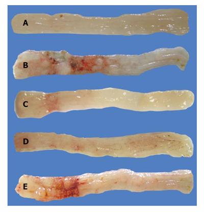
Figure 1 Macroscopic examination of the colonic mucosa before (A) and 24 h after acetic acid injection (B-E).
Mice were treated two days previously with (B) saline (damage score 4, square area of injury 40%); (C) prednisolone 5 mg/kg (damage score 2, square area of injury 1%); (D) OP, 100 mg/kg (damage score 2, square area of injury 3%); (E) apple pectin, 100 mg/kg (damage score 3, square area of injury 25%). The colon without the cecum was removed and opened along the mesenteric border.
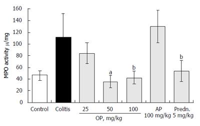
Figure 2 Effect of oxycoccusan OP on myeloperoxidase (MPO) activity in colonic tissue of mice with acetic acid-induced colitis.
Values are means ± SD (n = 7), aP < 0.05, bP < 0.01 vs the colitis group.
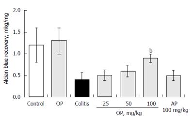
Figure 3 Effect of oxycoccusan OP (100 mg/kg) on the levels of bound mucus in the colon of mice.
Values are means ± SD (n = 7), bP < 0.01 vs the appropriate control.
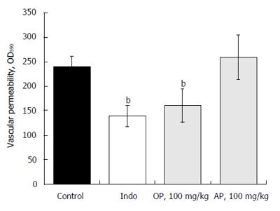
Figure 4 Effect of oxycoccusan OP on the vascular permeability of the healthy mice.
Values are means ± SD (n = 7), bP < 0.01 vs the control.
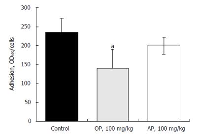
Figure 5 Effect of oxycoccusan OP on the adhesion of peritoneal leukocytes of the healthy mice.
Values are means ± SD (n = 7), aP < 0.05 vs the control.
- Citation: Popov SV, Markov PA, Nikitina IR, Petrishev S, Smirnov V, Ovodov YS. Preventive effect of a pectic polysaccharide of the common cranberry Vaccinium oxycoccos L. on acetic acid-induced colitis in mice. World J Gastroenterol 2006; 12(41): 6646-6651
- URL: https://www.wjgnet.com/1007-9327/full/v12/i41/6646.htm
- DOI: https://dx.doi.org/10.3748/wjg.v12.i41.6646


