Copyright
©2006 Baishideng Publishing Group Co.
World J Gastroenterol. Oct 28, 2006; 12(40): 6564-6566
Published online Oct 28, 2006. doi: 10.3748/wjg.v12.i40.6564
Published online Oct 28, 2006. doi: 10.3748/wjg.v12.i40.6564
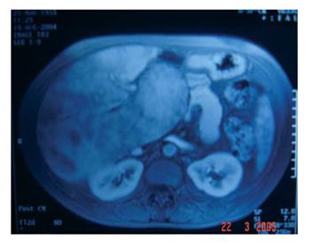
Figure 1 MRI in arterial phase showing a massive hepatomegaly with enlarged caudate lobe and patchy enhancement of contrast.
A branch of portal vein is shown (arterio-portal shunt).
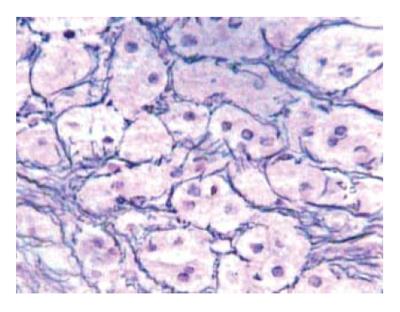
Figure 2 Liver biopsy reveals perisinusoidal and pericellular fibrosis (Gomori¡'s reticulin stain, original magnification x 100).
- Citation: Robles-Medranda C, Lukashok H, Biccas B, Pannain VL, Fogaça HS. Budd-Chiari like syndrome in decompensated alcoholic steatohepatitis and liver cirrhosis. World J Gastroenterol 2006; 12(40): 6564-6566
- URL: https://www.wjgnet.com/1007-9327/full/v12/i40/6564.htm
- DOI: https://dx.doi.org/10.3748/wjg.v12.i40.6564


