Copyright
©2006 Baishideng Publishing Group Co.
World J Gastroenterol. Oct 28, 2006; 12(40): 6458-6463
Published online Oct 28, 2006. doi: 10.3748/wjg.v12.i40.6458
Published online Oct 28, 2006. doi: 10.3748/wjg.v12.i40.6458
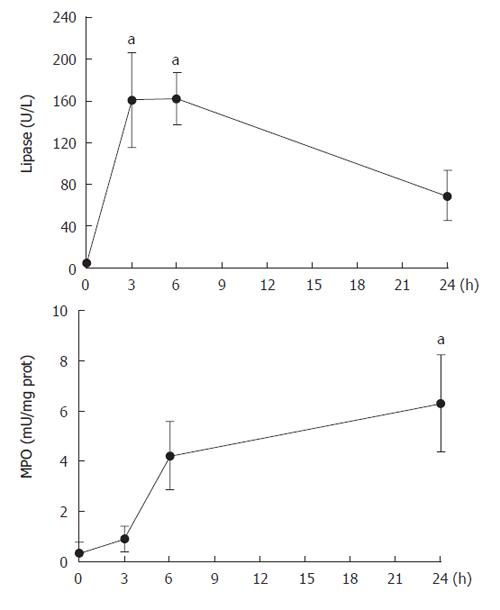
Figure 1 Plasma lipase and pancreas MPO activity after retrograde infusion of contrast media.
(aP < 0.05 vs t = 0).
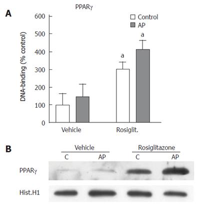
Figure 2 PPARγ activation.
A: PPARγ DNA binding activity of pancreas nuclear extracts expressed as % of control activity. No significant differences were observed on PPARγ binding to DNA after pancreatitis induction. By contrast, PPARγ binding to DNA is strongly induced by rosiglitazone in both control and pancreatitis groups. aP < 0.05 Rosiglitazone-treated vs vehicle-treated groups. B: Western blot of nuclear PPARγ confirmed the nuclear translocation of PPARγ protein after rosiglitazone administration in both control and pancreatitis groups. Western blot was representative of three different experiments.
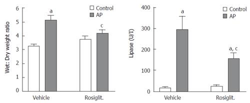
Figure 3 Pancreatic tissue edema and plasma lipase activity 6 h after contrast medium infusion.
Retrograde administration of contrast medium induced increases in edema and lipase activity. These increases were partially prevented by pre-treatment of rosiglitazone. aP < 0.05 AP vs their corresponding control; cP < 0.05 rosiglitazone-treated AP group vs vehicle-treated AP group.
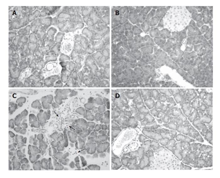
Figure 4 Histological examination of the pancreas (x 200).
A: Control pancreas showed normal acinar structure; B: No morphological changes were observed after rosiglitazone administration in control animals; C: Experimental ERCP-induced pancreatic damage reflected in interlobular and interacinar edema and areas of leukocyte infiltration (arrows). Acinar necrosis was not observed; D: Rosiglitazone pre-treatment before contrast medium infusion resulted in a reduced edema and absence of leukocyte cell infiltration.
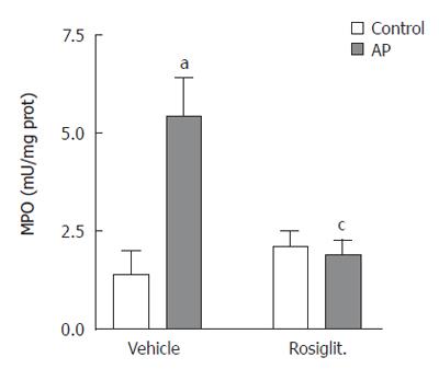
Figure 5 Myeloperoxidase activity in pancreas.
MPO activity was significantly increased in post-ERCP induced pancreatitis in comparison with vehicle. Rosiglitazone treatment prevented this increase. aP < 0.05 AP vs control; cP < 0.05 rosiglitazone-treated AP group vs Vehicle-treated AP group.
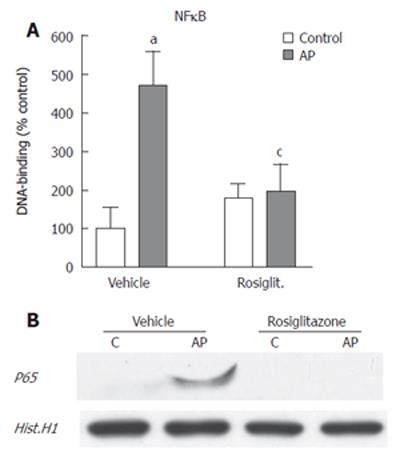
Figure 6 NFκB activation.
A: NFκB DNA binding activity of pancreas nuclear extracts expressed as % of control p65 activity. Post-ERCP induced pancreatitis resulted in increased levels of p65 binding activity. This increase was inhibited by rosiglitazone pre-treatment. aP < 0.05 AP vs Control; cP < 0.05 rosiglitazone-treated AP group vs vehicle-treated AP group; B: Western blot of nuclear p65 confirmed the nuclear translocation of p65 protein during ERCP-induced pancreatitis. This nuclear translocation was not observed after rosiglitazone administration. Western blot was representative of three different experiments.
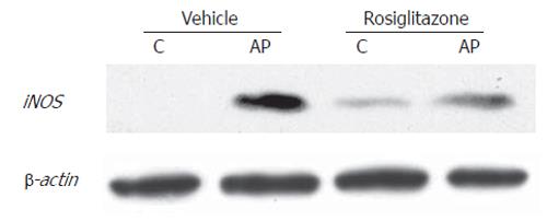
Figure 7 Inducible NO synthase (iNOS) expression.
Western blot of cytoplasmatic iNOS showed increased expression of this protein during post-ERCP induced pancreatitis. This increase was reduced by rosiglitazone pre-treatment. Results are representative of three separate experiments.
- Citation: Folch-Puy E, Granell S, Iovanna JL, Barthet M, Closa D. Peroxisome proliferator-activated receptor γ agonist reduces the severity of post-ERCP pancreatitis in rats. World J Gastroenterol 2006; 12(40): 6458-6463
- URL: https://www.wjgnet.com/1007-9327/full/v12/i40/6458.htm
- DOI: https://dx.doi.org/10.3748/wjg.v12.i40.6458


