Copyright
©2006 Baishideng Publishing Group Co.
World J Gastroenterol. Oct 21, 2006; 12(39): 6290-6298
Published online Oct 21, 2006. doi: 10.3748/wjg.v12.i39.6290
Published online Oct 21, 2006. doi: 10.3748/wjg.v12.i39.6290
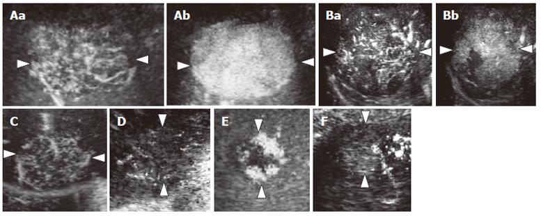
Figure 1 Diagram showing enhancement patterns of hepatic tumors in the arterial phase.
A: Intratumoral vessels in the early arterial phase (a) with homogeneous enhancement in the late arterial phase (b) (pattern A1); B: Intratumoral vessels in the early arterial phase (a) with heterogeneous enhancement in the late arterial phase (b) (pattern A1); C: Intratumoral vessels without homogeneous or heterogeneous enhancement (pattern A2); D: Peritumoral vessels alone (pattern A3); E: Peripheral nodular enhancement without tumor vessels (pattern A4); F: No enhancement and no tumor vessels (pattern A5).
Figure 2 Diagram showing enhancement patterns of hepatic tumors in the portal phase.
A: Homogeneous enhancement (pattern P1); B: Heterogeneous enhancement (pattern P1); C: Perfusion defect (pattern P2); D: Ring enhancement (pattern P3); E: Peripheral nodular enhancement (pattern P4).
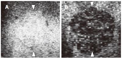
Figure 3 Diagram showing enhancement patterns of hepatic tumors in the late phase.
A: Lso-echoic (pattern L1); B: Hypo-echoic (pattern L2).
- Citation: Numata K, Isozaki T, Morimoto M, Sugimori K, Kunisaki R, Morizane T, Tanaka K. Prospective study of differential diagnosis of hepatic tumors by pattern-based classification of contrast-enhanced sonography. World J Gastroenterol 2006; 12(39): 6290-6298
- URL: https://www.wjgnet.com/1007-9327/full/v12/i39/6290.htm
- DOI: https://dx.doi.org/10.3748/wjg.v12.i39.6290


