Copyright
©2006 Baishideng Publishing Group Co.
World J Gastroenterol. Oct 21, 2006; 12(39): 6280-6284
Published online Oct 21, 2006. doi: 10.3748/wjg.v12.i39.6280
Published online Oct 21, 2006. doi: 10.3748/wjg.v12.i39.6280
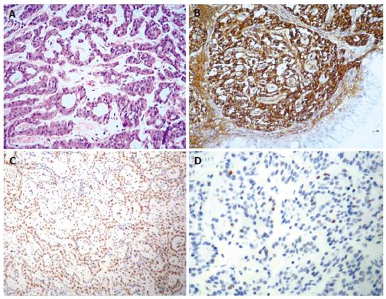
Figure 1 Histochemical staining.
A: Tumor cells arranged in ribbons, tubular structures or small nests (HE x 20); B: Chromogranin immunoexpression (x 10); C: p27 immunoexpression (x 20); D: p21 immunoexpression (x 20).
- Citation: Doganavsargil B, Sarsik B, Kirdok FS, Musoglu A, Tuncyurek M. p21 and p27 immunoexpression in gastric well differentiated endocrine tumors (ECL-cell carcinoids). World J Gastroenterol 2006; 12(39): 6280-6284
- URL: https://www.wjgnet.com/1007-9327/full/v12/i39/6280.htm
- DOI: https://dx.doi.org/10.3748/wjg.v12.i39.6280


