Copyright
©2006 Baishideng Publishing Group Co.
World J Gastroenterol. Oct 14, 2006; 12(38): 6229-6231
Published online Oct 14, 2006. doi: 10.3748/wjg.v12.i38.6229
Published online Oct 14, 2006. doi: 10.3748/wjg.v12.i38.6229
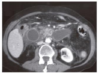
Figure 1 Dilated jejunal loop crossing the midline.
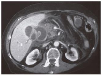
Figure 2 The dilated afferent loop and the stone (arrow).
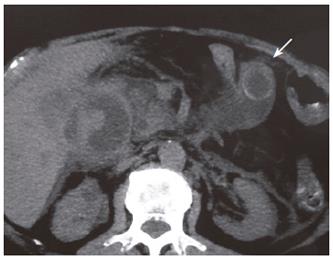
Figure 3 Stone in more detail (arrow).
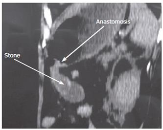
Figure 4 The relation between the gastrojejunal anastomosis (right arrow) and the stone (left arrow) in a tomographic reconstruction.
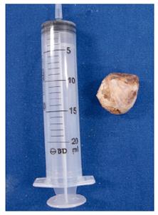
Figure 5 Biliary stone (with a diameter of 3 centimeters) after endoscopic removal.
- Citation: Dias AR, Lopes RI. Biliary stone causing afferent loop syndrome and pancreatitis. World J Gastroenterol 2006; 12(38): 6229-6231
- URL: https://www.wjgnet.com/1007-9327/full/v12/i38/6229.htm
- DOI: https://dx.doi.org/10.3748/wjg.v12.i38.6229


