Copyright
©2006 Baishideng Publishing Group Co.
World J Gastroenterol. Oct 14, 2006; 12(38): 6182-6187
Published online Oct 14, 2006. doi: 10.3748/wjg.v12.i38.6182
Published online Oct 14, 2006. doi: 10.3748/wjg.v12.i38.6182
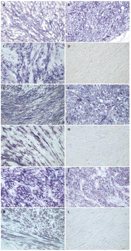
Figure 1 Immunohistochemical staining reveals VEGF expression in the cytoplasm of GIST (A), leiomyoma (B) and schwannoma (C) cells; VEGFR-1 expression in the membrane and cytoplasm of GIST (E), leiomyoma (F) and schwannoma (G) cells; VEGFR-2 expression in the membrane and cytoplasm of GIST (I), leiomyoma (J) and schwannoma (K) cells; negative staining of GIST for VEGF, VEGFR-1 or VEGFR-2 in Figure 1D, H or L, respectively.
BCIP/NBT reaction product demonstrating VEGF, VEGFR-1 and 2 levels. (magnification: x 200).
- Citation: Nakayama T, Cho YC, Mine Y, Yoshizaki A, Naito S, Wen CY, Sekine I. Expression of vascular endothelial growth factor and its receptors VEGFR-1 and 2 in gastrointestinal stromal tumors, leiomyomas and schwannomas. World J Gastroenterol 2006; 12(38): 6182-6187
- URL: https://www.wjgnet.com/1007-9327/full/v12/i38/6182.htm
- DOI: https://dx.doi.org/10.3748/wjg.v12.i38.6182


