Copyright
©2006 Baishideng Publishing Group Co.
World J Gastroenterol. Oct 14, 2006; 12(38): 6172-6177
Published online Oct 14, 2006. doi: 10.3748/wjg.v12.i38.6172
Published online Oct 14, 2006. doi: 10.3748/wjg.v12.i38.6172
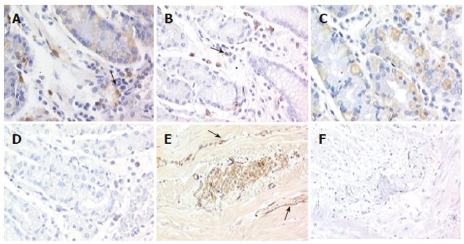
Figure 1 Expression of Kit in normal gastric tissue and gastroparesis (x 200).
In both normal (A) and gastroparetic stomach (B), Kit was detected on mast cells (arrows) used as an internal control; In normal gastric samples, positivity is shown on parietal cells (C) and at the intrinsic innervation level (E, arrows), whereas in gastroparesis no staining is present at either levels (D, F).
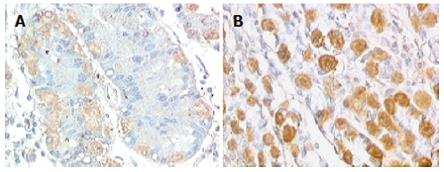
Figure 2 Expression of SCF in normal gastric tissue (A) and gastroparesis (B) (x 200).
Intense intracytoplasmatic expression of SCF with membrane reinforcement in gastroparesis parietal cells was present.
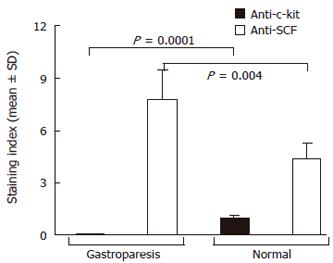
Figure 3 Quantitative evaluation of SCF expression on parietal cells in gastric tissue specimens from control subjects and the patient with gastroparesis.
Differences in the immunoreactive score, obtained as described in the text, were assessed by t-test.
- Citation: Battaglia E, Bassotti G, Bellone G, Dughera L, Serra AM, Chiusa L, Repici A, Mioli P, Emanuelli G. Loss of interstitial cells of Cajal network in severe idiopathic gastroparesis. World J Gastroenterol 2006; 12(38): 6172-6177
- URL: https://www.wjgnet.com/1007-9327/full/v12/i38/6172.htm
- DOI: https://dx.doi.org/10.3748/wjg.v12.i38.6172


