Copyright
©2006 Baishideng Publishing Group Co.
World J Gastroenterol. Oct 14, 2006; 12(38): 6156-6160
Published online Oct 14, 2006. doi: 10.3748/wjg.v12.i38.6156
Published online Oct 14, 2006. doi: 10.3748/wjg.v12.i38.6156
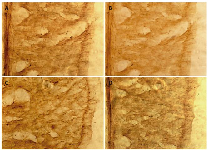
Figure 1 Microscopic photography of VIP-positive fibers of gastric smooth muscle in antrum (× 400).
A: Moderate immunoreactive staining of VIP-positive nerve fibers in control group (× 400); B: Weak immunoreactive staining of VIP-positive nerve fibers in model group (× 400); C: Strong immunoreactive staining of VIP-positive nerve fibers in EA group (× 400); D: Moderate immunoreactive staining of VIP-positive nerve fibers in non-acupoint group (× 400).
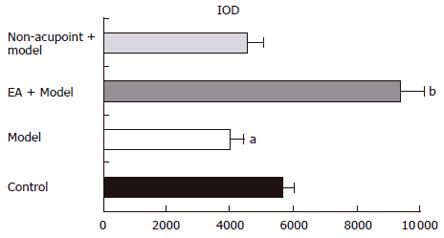
Figure 2 IOD analysis of the effect of EA on VIP immunoreactivity.
aP < 0.05 vs control group; bP < 0.01 vs model group. Data are mean ± SD, n = 10.
- Citation: Shen GM, Zhou MQ, Xu GS, Xu Y, Yin G. Role of vasoactive intestinal peptide and nitric oxide in the modulation of electroacupucture on gastric motility in stressed rats. World J Gastroenterol 2006; 12(38): 6156-6160
- URL: https://www.wjgnet.com/1007-9327/full/v12/i38/6156.htm
- DOI: https://dx.doi.org/10.3748/wjg.v12.i38.6156


