Copyright
©2006 Baishideng Publishing Group Co.
World J Gastroenterol. Sep 21, 2006; 12(35): 5699-5704
Published online Sep 21, 2006. doi: 10.3748/wjg.v12.i35.5699
Published online Sep 21, 2006. doi: 10.3748/wjg.v12.i35.5699
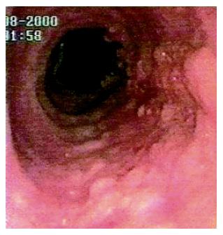
Figure 1 A sessile villous-like polyp extending for the whole length of BE.
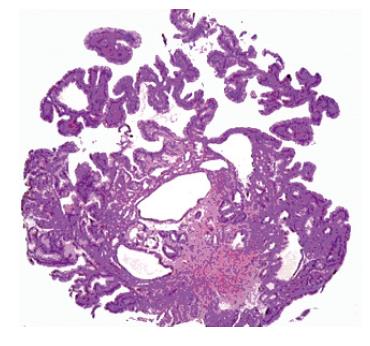
Figure 2 Hyperplastic polyp from the gastroesophageal junction was comprised of cardiac type mucosa with foveolar hyperplasia and cystic dilatation of gastric pits (HE x 100).
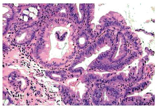
Figure 3 Sessile lesion arising in the Barrett’s esophagus: the adenomatous villous-type polyp is composed of low grade dysplastic epithelium (HE x 250).
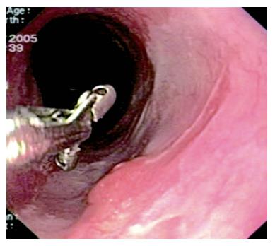
Figure 4 Areas of neosquamous re-epithelialization coexisting with tiny mucosal abnormalities.
- Citation: Ceglie AD, Lapertosa G, Blanchi S, Muzio MD, Picasso M, Filiberti R, Scotto F, Conio M. Endoscopic mucosal resection of large hyperplastic polyps in 3 patients with Barrett’s esophagus. World J Gastroenterol 2006; 12(35): 5699-5704
- URL: https://www.wjgnet.com/1007-9327/full/v12/i35/5699.htm
- DOI: https://dx.doi.org/10.3748/wjg.v12.i35.5699


