Copyright
©2006 Baishideng Publishing Group Co.
World J Gastroenterol. Sep 21, 2006; 12(35): 5674-5679
Published online Sep 21, 2006. doi: 10.3748/wjg.v12.i35.5674
Published online Sep 21, 2006. doi: 10.3748/wjg.v12.i35.5674
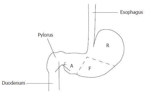
Figure 1 Different histological regions of rat stomach.
R: Cardic region or cardia; F: Fundic region or fundus; A: Pyloric region or antrum.
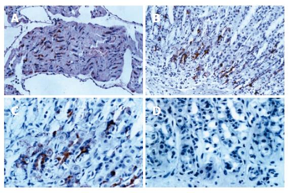
Figure 2 A: As a positive control, atrial myocytes show intense positive cytoplasmic staining for ANP (IHC × 200); B: Positive staining for ANP is localized to cytoplasm of mucosal cells in cardiac glands (IHC × 200); C: Positive staining for ANP is localized to cytoplasm of mucosal cells in cardiac glands (IHC × 400); D: As a negative control, complete absence of staining is observed when normal rabbit serum is substituted for ANP (IHC × 400).
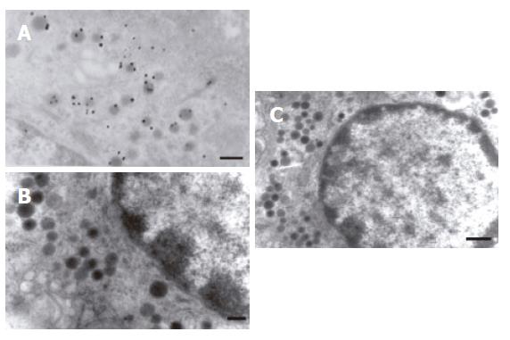
Figure 3 A: localization of immunogold labeled in the endocrine granule of the enterochromaffin cell (TEM × 20 000, bar = 200 nm); B: Negative control,complete absence of immunogold labeled in the endocrine granule of enterochromaffin cells when normal rabbit serum was substituted for anti-ANP antiserum (TEM × 20 000, Bar = 200 nm); C: Normal enterochromaffin cells in gastric mucosa (TEM × 15 000, Bar = 500 nm).
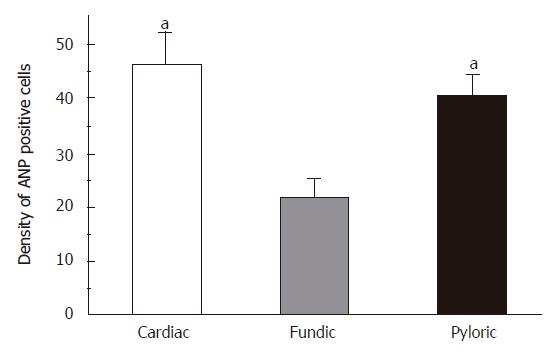
Figure 4 The distribution of ANP synthesizing cells in different regions of stomach in rats.
Mean ± SD. aP < 0.05 vs fundic region.
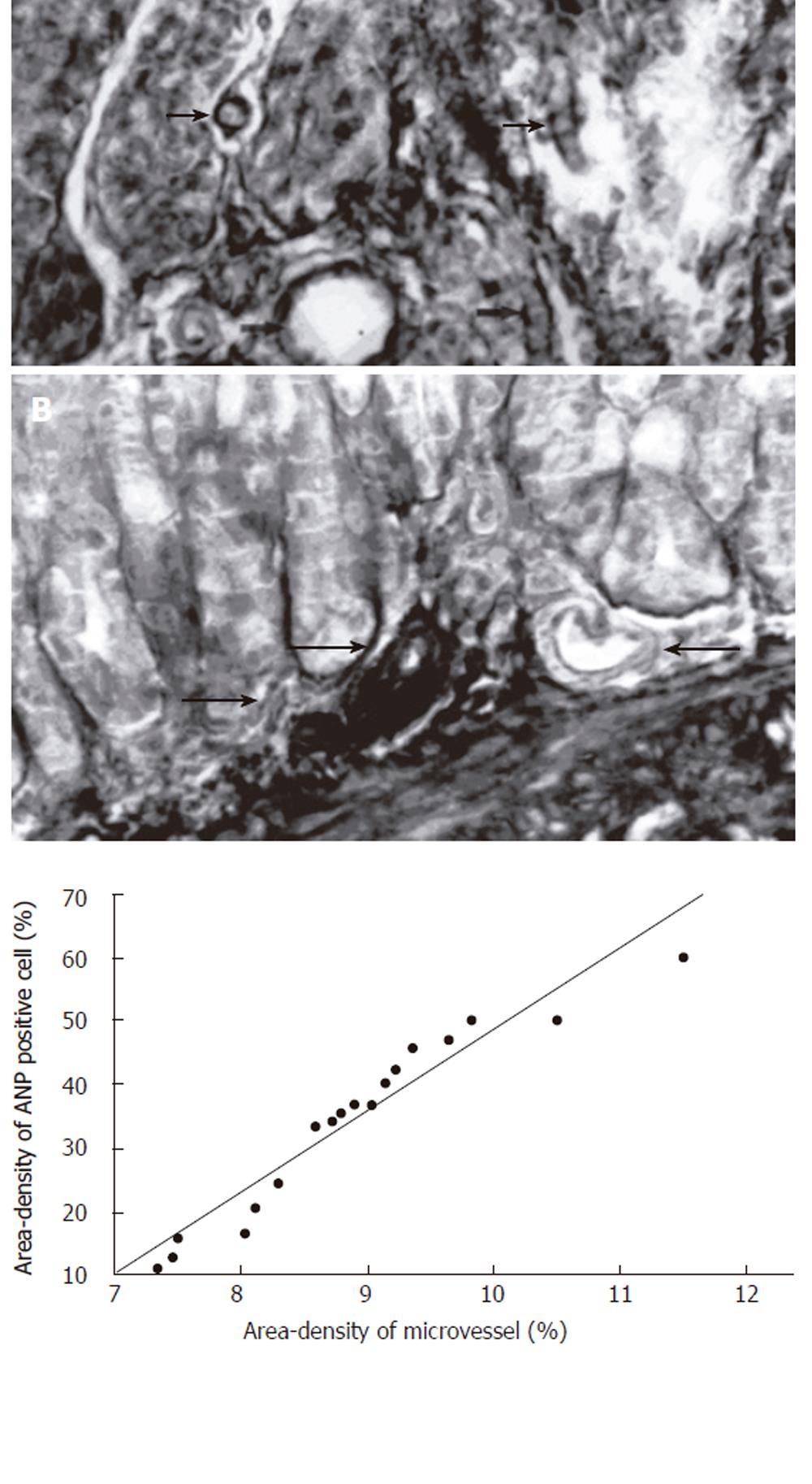
Figure 5 A: Microvessels of gastric mucous were in wriggled way and cut into different cross-sections, it could be clearly observed in distinct three-dimensions (arrow) (TA-Fe × 400); B: Some branch arteries from the large vessels run into the basal glands of the gastric mucosa (arrow) (TA-Fe × 400); C: There is positive significant relationship between the positive rate (%) of ANP-synthesizing cells and microvessel density (%) in cardiac region mucosa of rats (r = 0.
53, P < 0.05, n = 18).
- Citation: Li CH, Pan LH, Li CY, Zhu CL, Xu WX. Localization of ANP-synthesizing cells in rat stomach. World J Gastroenterol 2006; 12(35): 5674-5679
- URL: https://www.wjgnet.com/1007-9327/full/v12/i35/5674.htm
- DOI: https://dx.doi.org/10.3748/wjg.v12.i35.5674


