Copyright
©2006 Baishideng Publishing Group Co.
World J Gastroenterol. Sep 14, 2006; 12(34): 5562-5564
Published online Sep 14, 2006. doi: 10.3748/wjg.v12.i34.5562
Published online Sep 14, 2006. doi: 10.3748/wjg.v12.i34.5562
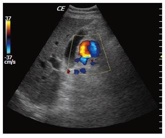
Figure 1 Color –Doppler US showed an anechoic mass, 2.
5 cm in diameter, next to the head of the pancreas with turbulent flow inside.
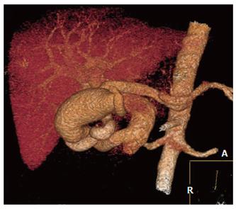
Figure 2 MDCT 3D reconstruction showed the presence of a large aneurysm filled with a dilated gastroduodenal artery and draining into the superior mesenteric vein.
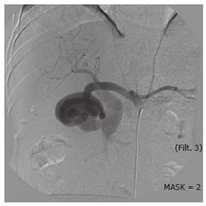
Figure 3 Celiac axis arteriogram confirmed a large aneurysm of gastro-duodenal artery and a fistulous communication with the main portal vein.
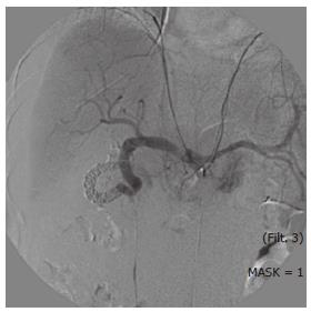
Figure 4 Arteriogram performed after coils embolization showed complete occlusion of the fistula.
- Citation: Marrone G, Caruso S, Miraglia R, Tarantino I, Volpes R, Luca A. Percutaneous transarterial embolization of extrahepatic arteroportal fistula. World J Gastroenterol 2006; 12(34): 5562-5564
- URL: https://www.wjgnet.com/1007-9327/full/v12/i34/5562.htm
- DOI: https://dx.doi.org/10.3748/wjg.v12.i34.5562


