Copyright
©2006 Baishideng Publishing Group Co.
World J Gastroenterol. Sep 7, 2006; 12(33): 5396-5398
Published online Sep 7, 2006. doi: 10.3748/wjg.v12.i33.5396
Published online Sep 7, 2006. doi: 10.3748/wjg.v12.i33.5396
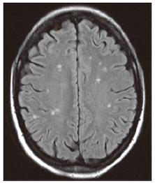
Figure 1 MRI (T2w/FLAIR) of the centrum semiovale showing multiple foci of subcortical signal enhancements which are characteristic but not specific for vasculitis.
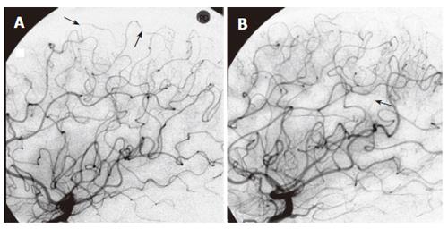
Figure 2 Left internal carotid artery (ACI) demonstrating irregularities of the distal parts of branches of the callosomarginal artery (arrows) (A) and right ACI demonstrating the break of a central branch of the middle cerebral artery (arrow) (B).
- Citation: Lüth S, Birklein F, Schramm C, Herkel J, Hennes E, Müller-Forell W, Galle P, Lohse A. Multiplex neuritis in a patient with autoimmune hepatitis: A case report. World J Gastroenterol 2006; 12(33): 5396-5398
- URL: https://www.wjgnet.com/1007-9327/full/v12/i33/5396.htm
- DOI: https://dx.doi.org/10.3748/wjg.v12.i33.5396


