Copyright
©2006 Baishideng Publishing Group Co.
World J Gastroenterol. Sep 7, 2006; 12(33): 5379-5383
Published online Sep 7, 2006. doi: 10.3748/wjg.v12.i33.5379
Published online Sep 7, 2006. doi: 10.3748/wjg.v12.i33.5379
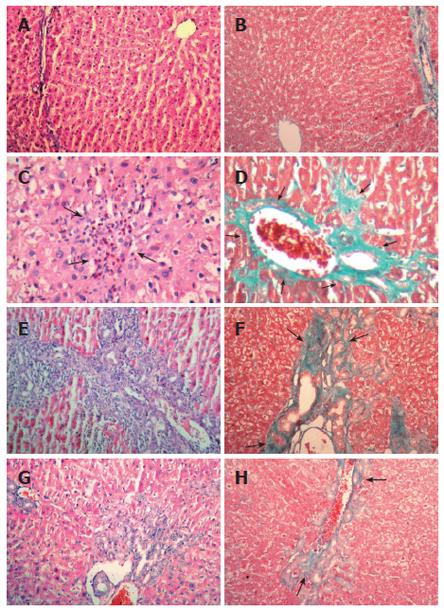
Figure 1 Morphological changes in the livers of different group rats (HE × 200; Masson’s trichrome × 200).
No morphological damage was observed in any of the rats in the sham-control group (A and B). In the untreated group, proliferation of portal and periportal biliary ductules with disorganization of the hepatocytes plates, dilated portal spaces and areas of polymorphonuclear leukocyte infiltrate, hepatocytes necrosis and fibrosis were observed (C and D). Moderate hepatocytes necrosis and fibrosis were present in the low-dose dexa group (E and F). The high-dose dexa group showed a remarkably less necrosis and fibrosis (G and H). A, C, E and G are stained with HE; B, D, F and H are stained with Masson’s trichrome.
- Citation: Eken H, Ozturk H, Ozturk H, Buyukbayram H. Dose-related effects of dexamethasone on liver damage due to bile duct ligation in rats. World J Gastroenterol 2006; 12(33): 5379-5383
- URL: https://www.wjgnet.com/1007-9327/full/v12/i33/5379.htm
- DOI: https://dx.doi.org/10.3748/wjg.v12.i33.5379


